Choose timezone
Your profile timezone:
We extend our heartfelt thanks to everyone who contributed to the success of the 12th International Workshop on X-ray Radiation Damage to Biological Samples (RD12) at the Paul Scherrer Institute.
Your engagement—whether as a speaker, poster presenter, participant, sponsor, or organizer—was at the heart of what made RD12 such a vibrant and inspiring event. Over the course of three days, the exchange of ideas across disciplines, institutions, and countries created a dynamic forum that truly advanced the field of X-ray radiation damage research.
We’re especially grateful to our invited speakers and poster contributors for sharing their latest findings, and to the Swiss Light Source and SwissFEL teams for the engaging facility tours. The workshop would not have been possible without the dedication of the organizing committee and the generous support of our sponsors.
We look forward to continuing this momentum through the upcoming special issue in Acta Crystallographica Section D, and we’re already excited for what RD13 will bring.
Thank you for being part of the RD12 community—your presence made it unforgettable.
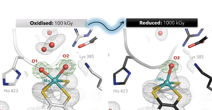
The molybdenum enzyme arsenite oxidase (Aio), is a promising biocatalyst for the detoxification and biosensor applications [1]. So far, its structural characterization and complete reaction mechanism understanding has been limited to artifactually reduced states of the Mo cofactor (Moco) caused by X-ray photoreduction (PDB 1g8k, 4aay) [2-4].
Here, we present the first crystallographic evidence of X-ray-induced photo-reduction in Alcaligenes faecalis Aio, tracking active-site geometric changes across incremental radiation doses (100–1000 kGy) at the PETRA III P14 beamline. High-resolution structures (1.5 Å) reveal that photo-reduction triggers the loss of a labile oxo ligand (O1), while displacing the Mo atom toward the dithiolene plane.
Low-dose data (100 kGy) allowed us to capture, for the first time, the Mo(VI) centre in a six-coordinated geometry with asymmetric cis-dioxo coordination (Mo–O1: 1.8 Å; Mo–O2: 2.1 Å). Reduction of the Moco also flattened the twist angle of the pterin from 31° to 17°, modulating the active site catalysis.
Our work demonstrates that radiation damage artifacts—prevalent in metalloenzyme crystallography—can obscure mechanistic insights. These findings underscore the need for dose-optimized structural data to refine AI-driven models of metalloenzyme mechanisms and advance the rational engineering of Aio for biosensing and bioremediation.
References
[1] Male, K.B. et al. (2007) Anal. Chem, 79(20), 7831–7837.
[2] Warelow T.P. et al., (2013) PLoS One, 8(8): e72535.
[3] Ellis, P.J. et al. (2001) Structure, 9(2), 125-132.
[4] Engrola, F. et al. (2023) J. Biol. Chem, 299(8), 105036.
Radiation damage is a faithful attender to X-ray crystallographic studies of metallo-proteins. Thus, care is taken to limit the effects of radiation damage and avoid consequent misinterpretation of X-ray crystallographic results. In our study, we applied defined doses to selectively probe the redox states involved in metal binding.
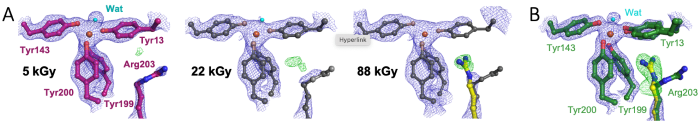
We studied the cyanobacterial iron binding protein FutA, an ABC transporter substrate binding protein that can also act as an intracellular iron binding protein [1]. We determined crystallographic structures using X-ray and neutron radiation characterised as ferrous [Fe(II)] and ferric [Fe(III)] complexes [2]. These states are distinguished by protein conformation, particularly the positioning of the positively charged Arg203 side chain as part of the iron binding site in the [Fe(II)] complex.
.png)
We captured the transition between [Fe(III)] and [Fe(II)] states upon X-ray photoreduction with a dose series using a serial synchrotron crystallography fixed target approach, see panel A. Using a novel XFEL X-ray pump-probe approach, we uncovered how Arg203 functions as a molecular switch, enabling accommodation of different iron oxidation states, see panel B [2]. The switching capability of the single FutA protein provides functional insight and suggests genome streamlining, where the loss of specialised FutA variants may reflect ecological adaptation.
References
[1] Polyviou, D. et al. (2018) J. Biol. Chem., 293, 18099-18109.
[2] Bolton, R. et al. (2024) PNAS, 121, e2308478121.
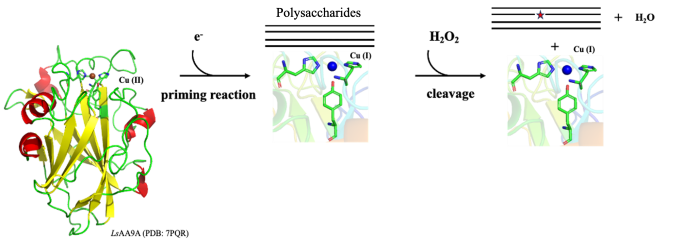
Lytic polysaccharide monooxygenases are enzymes [1] binding their active-site copper through the characteristic His-brace motif shown above including two His – one N-terminal – and often also a Tyr. The reaction cycle starts with reduction of the resting state Cu(II) to Cu(I) – in the laboratory usually using ascorbate as small molecule reductant. Despite the name, most LPMOs prefer hydrogen peroxide as co-substrate, to subsequently oxidatively cleave the glycosidic bonds in saccharides.
We have previously – through crystal cryo-structures of the model enzyme LsAA9A determined at high and low X-ray doses, and based on the hypothesis that X-ray induced photoreduction mimics natural priming reaction - reconstructed possible changes in geometry during the catalytic cycle and identified a small shortening of the Cu(II)-Tyr distance [2].
Aside from uncertainty on the biological significance of such shortening [3], we wanted to address concerns regarding the ability of macromolecular crystallography to reliably detect differences in the order of 0.1-0.2 Å. We thus carried out additional triplicate independent structure determination representing Cu(I)/Cu(II) states with/without the model substrates cellotriose, showing statistically significant differences only for the Cu(II)-Tyr distance with/without saccharide, but no other Cu-protein distance.
In order to assess whether additional general X-ray damage obscures similar shortening in the Cu(I) state induced by photoreduction, we are now comparing with cryo data collected after priming by chemical reduction with ascorbate at low X-ray doses.
Finally since X-ray-induced photoreduction of the active-site copper may closely approximate the chemical priming reaction, it holds potential as a trigger for time-resolved studies. To explore this possibility further, we are currently investigating the photoreduction process at room temperature.
References
[1] Tandrup, T., Frandsen, K.E.H., Johansen, K.S., Berrin, J.-G., Lo Leggio, L. (2018) Biochem. Soc. Trans. 46, 1431–1447
[2] Tandrup, T., Muderspach, S.J., Banerjee, S., Santoni, G., Ipsen, J.Ø., Hernández-Rollán, C., Nørholm, M.H.H., Johansen, K.S., Meilleur, F., Lo Leggio, L. (2022) IUCR Journal 9, 666-681.
[3] Wieduwilt, Lo Leggio and Hedegård (2024), Dalton Trans, 53, 5796-5807.
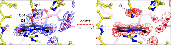
Urate oxidase (UOX) is the archetypal cofactor-independent oxidase, catalysing the O₂-mediated degradation of uric acid in many organisms. Previously, we demonstrated that the first step of this reaction is a dioxygenation reaction, forming 5-peroxyisourate (5-PIU) as an intermediate [1]. We also showed that 5-PIU is highly sensitive to radiation: during conventional single-axis X-ray diffraction experiments at 100 K, doses between 50-100 kGy caused visible damage to the C5–Op1 bond, and doses around 200 kGy led to complete bond rupture, accompanied by in situ release of O₂ [1,2]. Surprisingly, a subsequent synchrotron serial crystallography (SSX) experiment at room temperature (RT) showed no damage to this bond, even at an estimated dose of up to 70 kGy [3]. In this talk, I will discuss our more recent experiments exploring radiation-induced bond rupture under various experimental conditions—including gaussian vs. top-hat beam profiles, cryogenic vs. room temperature, and serial vs. conventional crystallography—in an effort to better connect these observations to the reaction mechanism.
.png)
References
[1] Bui S, von Stetten D, Jambrina PG, Prangé T, Colloc'h N, de Sanctis D, Royant A, Rosta E, Steiner RA (2014), Angew Chem Int Ed, 53, 13710-13714.
[2] Bui S and Steiner RA (2017) Curr Opin Struct Biol, 41,109-118.
[3] Zielinski KA, Prester A, Andaleeb H, Bui S, Yefanov O, Catapano L, Henkel A, Wiedorn MO, Lorbeer O, Crosas E, Meyer J, Mariani V, Domaracky M, White TA, Fleckenstein H, Sarrou I, Werner N, Betzel C, Rohde H, Aepfelbacher M, Chapman HN, Perbandt M, Steiner RA, Oberthuer D (2022) IUCrJ, 31:778-791.
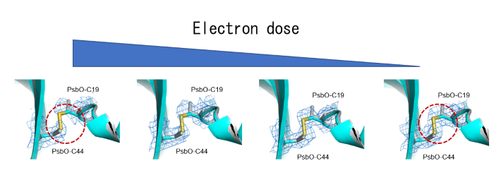
Photosystem II (PSII) plays a critical role in water-splitting and oxygen evolution. X-ray crystallography has elucidated its atomic structure and structures of several intermediate states. However, these structures are in the crystalline state, and the structure of the final state remains unresolved. In this study, we analyzed the structure of PSII in solution at a resolution of 1.95 Å using single-particle cryo-electron microscopy (cryo-EM). The obtained structure is similar to the crystal structure, but regions susceptible to redox state changes exhibited electron beam damages at high doses. By reducing the number of movie frames of electron micrographs from 50 to 2 to lower the beam dose, the damages were minimized while the resolution was comparable (Fig. a). I will discuss the details of the electron beam-induced damages and their minimization in this presentation.
.png)
References
[1] Kato K. et al. (2021) Commun Biol., 4, 382.
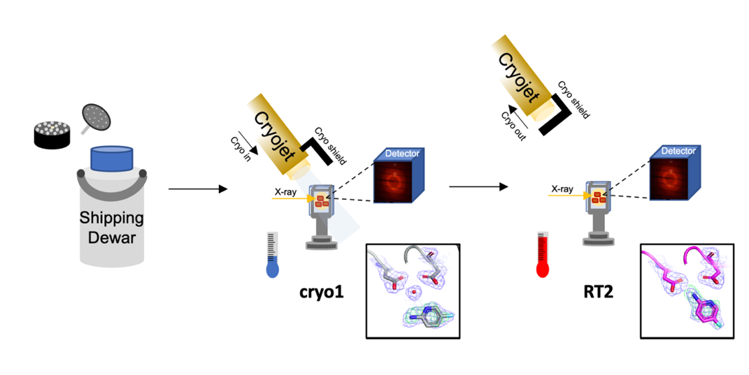
Cryogenic temperatures may introduce artefacts that limit the understanding of protein dynamics, crucial to their biological functions. To address this, we developed a room-temperature (RT) X-ray crystallographic method that captures movie-like structural snapshots triggered by temperature [1]. This method revealed binding-mode changes of TL00150, a 175.15 Da fragment, in endothiapepsin. Building on this, we further developed Cryo2RT, a high-throughput RT data-collection method using cryo-cooled crystals, which leverages the cryo-crystallography workflow [2]. This method has been applied to endothiapepsin with four soaked fragments, thaumatin, and SARS-CoV-2 3CLpro, Cryo2RT uncovered distinct ligand-binding modes at RT, not seen at cryogenic temperatures. To minimize radiation damage, X-ray doses were controlled below 500 kGy, a threshold considered safe for both cryo and RT crystallography. Despite similar doses, RT datasets showed slightly lower resolution and higher B-factors (30–40 Ų vs. ~20 Ų at cryo), likely due to increased atomic motion at RT. These findings provide insights into structural interpretation at RT and highlight Cryo2RT's potential for fragment-based screening and studying temperature-dependent dynamics.
References
[1] Huang, C.-Y., Aumonier, S., Engilberge, S., Eris, D., Smith, K. M. L., Leonarski, F., Wojdyla, J. A., Beale, J. H., Buntschu, D., Pauluhn, A., Sharpe, M. E., Metz, A., Olieric, V., and Wang, M. (2022) Acta Cryst. D78: 964-974. https://doi.org/10.1107/S205979832200612X
[2]: Huang, C.-Y., Aumonier, S., Olieric, V., and Wang, M. (2024) Acta Cryst. D80: 620-628. https://doi.org/10.1107/S2059798324006697