The 2nd UPLIFT School for “Artificial Intelligence in Radiotherapy”

→ Europe/Zurich
Congressi Stefano Franscini (CSF)

Congressi Stefano Franscini (CSF)

Ye Zhang (PSI - Paul Scherrer Institut), Antony John Lomax (PSI - Paul Scherrer Institut)
Description

The UPLIFT consortium (Upright Radiotherapy: Learning, Innovation, Fellowship and Training) is committed to advancing upright radiotherapy by addressing key scientific and clinical challenges, developing innovative solutions in imaging, treatment planning and workflows, and training the next generation of professionals. This initiative aims to revolutionise cancer treatment, making it more accessible, human-centred, and sustainable globally. 

As part of this mission, we proudly present the 2nd UPLIFT School: "Artificial Intelligence in Radiotherapy." This comprehensive six-day event explores the latest AI innovations transforming radio- and particle therapy.

2nd UPLIFT School –  Artificial Intelligence in Radiotherapy joint event with AI for Particle/Radio- Therapy (AI4PT|RT) symposium

Program Highlights

  • World-Class Faculty: Hear from 17 internationally renowned speakers who will deliver expert presentations on the frontiers of radiotherapy applications and cutting-edge AI development.
  • Joint AI4PT|RT Symposium (Days 5, 6 & 7): The event culminates in a 2.5-day symposium on AI for Particle/Radio-Therapy, highlighting high-impact scientific contributions that leverage AI to enhance radiotherapy.
  • Open Scientific Presentations (Days 6 & 7): To showcase the latest research from across the community, more than 10 oral presentation slots have been exclusively reserved for open scientific submissions, selected through a competitive abstract evaluation process.
  • Poster Sessions & Travel Grants: Two interactive poster sessions provide an additional platform for early-stage researchers (ESRs) and students to present their work. To support and encourage broader participation, three travel grants will be awarded to the authors of the top-ranked abstracts.

  • Hands-On AI Academy (Days 1 & 2): Practical training sessions designed to equip beginner developers with essential AI skills tailored specifically for radiotherapy.
  • Career Development & Mentorship: Beyond the scientific presentations, dedicated mentorship sessions are integrated throughout the program to support the professional growth and career development of students and ESRs.
  • Alpine Networking (Day 2): The "Hike and Think Trail" in the Swiss Alps will provide attendees with a unique and inspiring setting to connect, collaborate, and network.

By seamlessly combining technical education, expert mentorship, and dynamic networking, the 2nd UPLIFT School is an essential experience for anyone focused on advancing the future of AI in radiotherapy and particle therapy.


UPLIFT school support team
    • 11:00
      Arrival @Monte Verità
    • 13:00
      Registration: UPLIFT school
    • 1
      Exercise I: Introduction to AI development platform and basic funtion
      Speaker: Dr Lennart Volz (GSI Helmholtzzentrum f¨ur Schwerionenforschung)
    • 2
      Welcome address
      Speakers: Mrs Therese Egli, Dr Ye Zhang (PSI - Paul Scherrer Institut)
    • 19:00
      Welcome dinner
    • 3
      Opening: 2nd UPLIFT school
      Speaker: Dr Ye Zhang (PSI - Paul Scherrer Institut)
    • 4
      Opening plenary: AI in RT

      Stine S Korreman, PhD, is Professor of Medical Physics at Aarhus University, Denmark.
      She leads a research group on AI for medical image analysis in radiotherapy with focus on segmentation and dose prediction, and translation of results from research to clinical practice.
      She is director of the research center AIM@CANCER: AI-driven trustworthy foundation Models for personalized and effective CANCER radiotherapy
      She is chair of the European Society for Radiotherapy and Oncology (ESTRO) focus group on AI in radiotherapy and director of the ESTRO course on AI in radiotherapy.

      Speaker: Prof. Stine Korreman (Aarhus University)
    • 5
      Auto contouring - clinical status and limitation

      Dr. Jintao Ren is a radiotherapy AI postdoc researcher at Aarhus University Hospital and Aarhus University, Denmark. He is working on deep learning for medical image segmentation, tumor delineation, and adaptive radiotherapy. His recent work includes CT/PET lesion segmentation and MRI-guided head-and-neck gross tumor volume segmentation. His research focuses on translating AI contouring methods into clinically useful, safe, and trustworthy radiotherapy workflows

      Speaker: Dr Jintao Ren (Aarhus University)
    • 10:30
      Coffee break
    • 6
      AI-based medical image segmentation

      Professor Mauricio Reyes began his academic journey in 2003 when he was awarded a French-Chilean scholarship to conduct his PhD studies at INRIA, France. His research work focused on developing AI technologies to improve the workflow for cancer patients.
      His PhD thesis resulted in the development of novel image reconstruction techniques for respiratory motion compensation during PET imaging. This research work significantly improved the assessment of treatment response in lung cancer patients, demonstrating the potential of medical image computing technologies applied to medicine.
      Early in his postdoctoral years, Professor Reyes developed innovative high-throughput computational analysis methodologies for in-silico orthopaedic implant design. This research work was highlighted by the Swiss National Science Foundation as an example of front-end Swiss research. He was able to further translate this technology, changing the way orthopedic implants are designed nowadays using computational tools. In 2008, he co-founded Crisalix, a successful Swiss company offering AI-based technologies for simulation of reconstructive breast surgery.
      His work in computational modeling continued towards the challenging aspects of modelling pathology. Through EU-funded projects, he started in 2010 to develop AI solutions for neuro-oncology, with a particular focus on automated brain tumor burden assessment. His seminal research work, led to be the recipient of the MICCAI Young Scientist Publication award in 2016.
      His translational research efforts led to the first-of-its-kind FDA-approved AI technology for brain tumor patients, and a Boston-based startup with a subsidiary branch in Switzerland to further develop AI technologies. Throughout his career, Professor Reyes has raised over 11M EUR acquired funds, over 400 authored articles, with over 28000 citations, and a H-index of 65. His work has pioneered in the important aspects of robust AI, and explainable AI (XAI), in the field of medical image analysis."

      Speaker: Prof. Mauricio Reyes (University of Bern)
    • 12:00
      Lunch
    • 7
      Scientific writing and peer review

      After obtaining his PhD from the University of Aberdeen in Scotland and working for 2 years as a clinical medical physicist in the UK, Tony Lomax moved to Switzerland in 1992 to work at the Center for Proton Therapy at the Paul Scherrer Institute as a clinical medical physicist and researcher. In 2007 he became a Titular Professor in the Physics Department at ETH Zurich. From 2010-2014, he was on the editorial board of ‘Physics in Medicine and Biology’ and remains on the international advisory board. He is a regular member of the scientific committees for ASTRO, ESTRO, AAPM and the ICCR and from 2015 - 2023 was the physics co-chair of the PTCOG Scientific Program Sub-committee. In 2023 he became an honorary member of the Swiss Association of Scientific Radiation Oncology (SASRO) and in 2024 was the recipient of the Robert Wilson Award for his achievements in particle therapy from the Particle Therapy Cooperative Group (PTCOG). He is the author or co-author of over 330 peer reviewed papers, 13 book chapters and more than 500 invited and proffered conference presentations. From August 2026, he moved to work for RaySearch Laboratories in Stockholm

      Speaker: Prof. Antony John Lomax (PSI - Paul Scherrer Institut)
    • 8
      Conflict resolution

      Dr Tracy Underwood is Head of Translational Research at Leo Cancer Care. She is also a UKRI Future Leaders Fellow and an Honorary Senior Research Fellow at University College London. In 2011, Tracy founded the International Upright Radiotherapy and Imaging Research Consortium, bringing together researchers and clinicians from across the world to accelerate progress in this emerging area. Previously, as a radiotherapy researcher she worked at some of the most innovative clinics and academic departments worldwide, including Massachusetts General Hospital / Harvard Medical School in Boston, IUCT Oncopole in Toulouse, the Christie NHS Foundation Trust, the University of Manchester, and the University of Oxford. Together with her collaborators she is currently researching many aspects of upright radiotherapy, including: immobilisation methods, patient perspectives, anatomical changes with body position and treatment planning. She likes to spend her free time on the beach or in the forest with her husband and two young kids.

      Speaker: Dr Tracy Underwoods (Leo Cancer Care & UCL)
    • 9
      Intellectual property and patents

      Erik Engwall is Head of Physics and Algorithms, leading a department of 45 developers within physics, optimization, image analysis and visualization. He holds a PhD in physics from Uppsala University, Sweden. Prior to his current role, he held the role as Chief Physicist, overseeing the development within the areas of dose computation and advanced planning tools for photons, electrons, protons, heavier ions and brachytherapy. He takes an active role in the RayStation steering committee to determine which functionalities to introduce in the clinical RayStation versions. His main areas of interest span from fully automated workflow to novel concepts in radiotherapy, like upright treatments and proton arc therapy.

      Speaker: Dr Erik Engwall (RaySearch Laboratories)
    • 15:15
      Coffee break
    • 10
      Exercise II: DL based image segementation // commerical software
      Speaker: Lennart Volz (GSI Helmholtzzentrum f¨ur Schwerionenforschung)
    • 11
      UPLIFT supervisor meeting
      Speaker: Prof. Christian Graeff (GSI Helmholtzzentrum f¨ur Schwerionenforschung)
    • 19:00
      Dinner
    • 12
      Adaptive radiotherapy - offline, online, real-time

      Barbara Knäusl is a senior post-doctoral research associate and clinical medical physicist at the Department of Radiation Oncology, Medical University of Vienna. She is PI of the interdisciplinary Christian Doppler Laboratory for image- and knowledge-driven precision radiation oncology, leading a group of four PhD students, one postdoc and two assistants.
      Her research focuses on adaptive radiotherapy across photon and particle therapy with emphasis on image guidance, deformable image registration, breathing motion management, 4D treatments and outcome analysis. She has been involved for ten years in related research at the MedAustron Center for Ion Therapy and Research, most recently as PI and partner in the H2020 RAPTOR and RAPTOR+ consortium on real-time adaptive particle therapy, and as vice-coordinator of the FWF docFunds connect network on preclinical ion beam research.
      She has long engaged with technology development now converging in AI-driven radiotherapy, including automated segmentation, synthetic image creation and workflow automation. As editor-in-chief of Physics and Imaging in Radiation Oncology, an active member of the ESTRO scientific council and physics committee, and supervisor of PhD and MSc students, she contributes to a broad spectrum of the medical physics developments in radiation oncology.

      Speaker: Prof. Barbara Knaeusl (Medical University of Vienna)
    • 13
      Re-irradiation: from previous dose to robust treatment optimisation

      Ane Appelt, PhD, is Professor of Clinical Medical Physics at Rigshospitalet and the Technical University of Denmark, and Visiting Professor at the University of Leeds, UK. A clinically trained medical physicist, her research focuses on the development and implementation of novel radiotherapy methods & technology, with reirradiation as a major research theme.
      Her work in reirradiation spans clinical practice, methodology and technical development, including deformable image registration, cumulative dose evaluation, radiobiological dose rescaling and treatment plan optimisation. She has worked closely with industry partners to translate developments into commercial treatment planning software. She founded the ESTRO Physics Working Group on Reirradiation, is a core member of the ESTRO Reirradiation Focus Group, and co-leads the international Reirradiation Collaborative Group (ReCOG). Her work has contributed to international consensus recommendations for reirradiation and to the development of multicentre clinical studies and prospective trials.

      Speaker: Prof. Ane Appelt (Rigshospitalet & Technical University of Denmark)
    • 10:30
      Coffee break
    • 14
      AI boost deformable image registration

      Eliana Vasquez Osorio is a Reader in Computational Radiotherapy at the University of Manchester and an Honorary Researcher at The Christie NHS Foundation Trust. She trained as a computer scientist at Universidad EAFIT in Medellín, Colombia, and completed her PhD in Medical Physics at Erasmus University Rotterdam, the Netherlands. Her research develops computational and AI methods to make radiotherapy safer, more personalised and better understood, with a particular focus on reirradiation. Her work combines medical image computing, image registration, dose mapping, uncertainty quantification, outcome modelling and causal inference, with a particular interest in the development and safe clinical translation of AI-based methods for radiotherapy.

      Speaker: Dr Eliana Vasquez Osorio (The University of Manchester)
    • 12:00
      Lunch
    • 13:00
      Social activity (hike and think)
    • 15
      MRgRT - clinical status and future direction

      Medical Physics research group leader, University Hospital for Radiation Oncology Tübingen
      Full professor for Biomedical Physics, University of Tübingen
      ERC starting grant 2013 Biologically individualized Radiotherapy (bio-iRT)
      Medical Physics Expert
      PhD in Medical Physics, University of Tübingen
      Diploma in Physics, University of Stuttgart and General Engineering, École Centrale Paris

      Speaker: Prof. Daniela Thorwarth (Eberhard Karls Universität Tübingen)
    • 16
      Treatment outcome modelling

      Professor Marianne Aznar is Professor of Radiation Oncology Physics at the University of Manchester and Honorary medical physicist at the Christie NHS Foundation Trust. She trained as a clinical physicist in Canada and Denmark and joined the Radiotherapy-Related Research Group in 2017.
      Her research focuses on the side-effects of cancer treatment, especially for patient groups who can expect a long life after their original cancer treatments (diagnoses include, e.g., Hodgkin Lymphoma, breast cancer and childhood cancer), and more recently, the application of artificial intelligence to radiotherapy practice. Her current projects include BRAINatomy, looking at cognition in survivors of childhood brain tumours (https://www.mcrc.manchester.ac.uk/brainatomy-2/), and SMILE which looking at facial deformation and dental toxicities in children treated with Proton Beam Therapy (https://www.childrenscancerresearchfund.co.uk/pioneering-research-for-children-with-head-neck-cancers/). Internationally, she is involved with the European Society of Radiotherapy and Oncology (ESTRO) where she serves as an elected board member and a teaching faculty member. She also represents radiotherapy professionals on the Cardio-Oncology Council of the European Society of Cardiology and contributed to the recent international guidelines for cardio-oncology.
      Professor Aznar has published extensively in the field of medical physics and radiotherapy and serves as the physics editor for Radiotherapy and Oncology.

      Speaker: Prof. Marianne Aznar (The University of Manchester)
    • 10:30
      Coffee break
    • 17
      Advanced image guidance and treatment verification in PT

      Katia Parodi is since 2012 full professor and Founding Chair of Medical Physics at the Physics Faculty of the Ludwig-Maximilian-University in Munich, where she initiated a specialization curriculum in Medical Physics within the MSc in Physics. She studied Physics and Medical Physics at the University of Genoa, Italy, received in 2004 her Ph.D. in Physics from the University of Dresden, Germany, and subsequently pursued postdoctoral research at Massachusetts General Hospital and Harvard Medical School in Boston, USA. In 2006 she returned to Germany as tenured scientist and group leader at the Heidelberg Ion Therapy Center, obtaining in 2009 her Habilitation from the Heidelberg University.
      Her research interests are in high-precision image-guided radiotherapy, with focus on advanced methods for delivery, imaging and in-vivo monitoring of pre-clinical and clinical ion therapy. For her work, reported in over 300 peer-reviewed publications, she received several recognitions, and an ERC Consolidator grant in 2016. In 2017-2018 she served as president of the German Society for Medical Physics. She is since 2021 the Editor-in-Chief of the journal Physics in Medicine and Biology, and since 2024 an AAPM Fellow. In 2026 she also received the Innovation of the Year award of the Italian Chamber of Commerce for Germany.

      Speaker: Prof. Katia Parodi (Ludwig-Maximilians-Universität München)
    • 12:00
      Lunch
    • 18
      Round table discussion: Research/Clinic/Industry
      Speakers: Prof. Aswin Hoffmann (Helmholtz-Zentrum Dresden-Rossendorf), Prof. Petra Trnkova (Czech Technical University in Prague)
    • 15:00
      Coffee break
    • Poster dissusion: 1 (10+2min per person x 10)
      • 19
        Development of a 4D MRI Framework for Characterising Abdominal Motion in Supine and Upright Positions

        Objective
        The upright position of patients is related to significant changes of geometry and spatial arrangement of abdominal organs relative to the conventional supine position. As respiration acts on this altered anatomy, posture and breathing jointly determine the resulting abdominal motion. So far, this posture-breathing interaction which is relevant for upright RT is insufficiently characterised [1]. This work presents a 4D MRI framework for characterising breathing-related abdominal organ motion in supine and upright positions.

        Methods
        A free-breathing 4D MRI acquisition protocol was implemented on the 0.5 T open MROPEN EVO system using a HASTE-CINE (Half-Fourier Acquisition Single-shot Turbo Spin Echo) sequence [2,4]. Healthy volunteers were imaged in supine and upright positions with a stack of sagittal slices from the spine to the right of the liver. An interleaved acquisition strategy was employed for approximately 10 minutes whereby a navigator slice through the diaphragm dome was acquired every second. All other slices were acquired sequentially in between to generate multiple positional updates of the complete volume with an interval of N slices times 1 second. Resolution and slice thickness reflect a balance of image sharpness and signal-to-noise ratio. Respiratory signals are extracted from the navigator slice using image-based template tracking of the liver dome boundary after initial localisation, providing a displacement-based surrogate for diaphragm motion [3]. Each volunteer was scanned in supine and sitting or standing position. Scans were repeated to simulate separate visits.

        Results
        Initial volunteer datasets show variation in breathing amplitude, frequency, and regularity between supine and upright acquisitions, together with clear posture-dependent differences in abdominal anatomy and organ shape. Liver-dome displacement ranged from 10.4 to 38.3 mm in the supine position and from 7.4 to 21.9 mm in the standing position. In a separate group scanned in the supine and sitting positions, the corresponding ranges were 8.3 to 11.1 mm and 8.8 to 11.7 mm, respectively. These values represent the 5th-to-95th percentile range of the tracked liver-dome position. The measurements demonstrate the feasibility of capturing breathing-related motion across different volunteers, acquisitions, and postures.

        Outlook and Discussion
        This work establishes the technical foundation for posture-dependent 4D MRI investigations within the UPLIFT project using an open MR scanner with 0.5 T field. Next steps include expanding the volunteer cohort, finalising the respiratory signal extraction pipeline, and defining quantitative motion metrics to compare supine and upright postures. The results indicate that a positional support or fixation of shoulders may become a potential requirement for extending further studies with multiple repetitions over several days, for example, to address deformation, motion reproducibility, and inter-session variability.

        References
        [1] Volz L, Korte J, Martire MC, et al. Opportunities and Challenges of Upright Patient Positioning in Radiotherapy. Physics in Medicine & Biology, 2024.
        [2] Liu Y, Chang Z, Czito B, Bashir M, Yin FF, Cai J. 4D-MRI Using Sagittal Acquisition and Body Area as Respiratory Surrogate. International Journal of Radiation Oncology Biology Physics, 2013.
        [3] Liu Y, Yin FF, Czito BG, Bashir MR, Palta M, Cai J. Retrospective Four-Dimensional Magnetic Resonance Imaging With Image-Based Respiratory Surrogate: A Sagittal-Coronal-Diaphragm Point of-Intersection Motion Tracking Method. Journal of Medical Imaging, 2017.
        [4] Aljaafari L, Bird D, Buckley DL, Al-Qaisieh B, Speight R. A Systematic Review of 4D Magnetic Resonance Imaging Techniques for Abdominal Radiotherapy Treatment Planning. Physics and Imaging in Radiation Oncology, 2024.

        Acknowledgements: The work was performed in scope of UPLIFT Project (EU grant agreementNo.101168955)

        Speaker: Ms Anna Sampanai (ASG Superconductors Spa and Department of Mathematics, University of Genoa, Genoa, Italy)
      • 20
        Energy Layer Filtration for Upright Static Carbon Arcs

        Objectives:
        Beam delivery time (BDT) is an important consideration in particle arc therapy, particularly for carbon-ion arcs delivered using synchrotrons. Energy layer filtration can reduce BDT, but the current MU-based filtration may not optimally balance delivery efficiency and treatment quality. This study investigates alternative energy layer filtration strategies for upright static carbon arcs and evaluates their impact on the trade-off between treatment quality and BDT.

        Methods:
        Treatment plans were initialized using multiple beam directions and energy layers according to treatment site and case complexity. Three patients (two H&N and one intracranial) were robustly optimized using all investigated filtration approaches. During optimization, energy layers were filtered to a predefined number, with the required layers removed in a single filtration step. In addition to the MU-based filtration implemented in RayStation, three alternative strategies were investigated. First, a saliency-based approach inspired by Optimal Brain Damage published by LeCun et al. (OBD) was used to rank energy layers according to their contribution to the optimization objective and remove the least salient layers. Second, Group Lasso regularization was used to promote layer-wise sparsity, with groups defined according to either layer MU or cumulative saliency. Third, a delivery-time-aware strategy was developed by combining a traveling salesman problem (TSP) optimization of the delivery path with iterative removal of contiguous groups of layers having a low MU- or saliency-to-delivery-time ratio. The delivery path was recomputed after each filtration step.

        Results:
        OBD-based filtration reduced the objective value by more than 10% compared with MU-based filtration, while increasing BDT by approximately 5%. Group Lasso in conjunction with OBD provided an additional 2–3% improvement in objective value with little additional BDT (both relative to OBD). TSP-based filtration showed potential to further reduce BDT compared to MU-based filtering, with reductions of close to 10% and a deterioration of similar magnitude in objective value.

        Outlook & Discussion:
        Alternative energy layer filtration strategies show potential to improve the balance between treatment quality and delivery efficiency in upright static carbon arc therapy. Saliency-based filtration improved the optimization objective relative to MU-based filtration, while delivery-time-aware filtration demonstrated the potential for substantial BDT reduction albeit with a reduction in plan quality. Future work will evaluate the clinical relevance and robustness of these approaches and investigate more computationally efficient implementations of delivery-time-aware filtration.

        Funded by EU GA No. 101168955

        Speaker: Wouter van Weerelt (RaySearch Laboratories & KTH Royal University of Technology)
      • 21
        Dosimetric trade-offs between ITV expansion and robust optimization for inter-fractional anatomical variation in upright carbon-ion radiotherapy for lung cancer

        Objective: To quantify inter-fractional tumor variation in upright lung cancer radiotherapy and compare fraction-level and accumulated target coverage and normal-tissue sparing across five ITV-based planning strategies.
        Methods: Eight patients with lung cancer underwent upright planning CT and serial daily in-room CT. Following rigid alignment, target contours were propagated to daily CTs by deformable image registration. Inter-fractional variation was quantified using Dice similarity coefficient (DSC), 95th-percentile Hausdorff distance (HD95), relative volume change (ΔV), and center-of-mass (COM) displacement. Five CIRT plans were generated for each patient: conventional optimization using 3-, 5-, or 7-mm ITV expansions (M3, M5, and M7), and ITV-based robust optimization with 3- or 5-mm setup uncertainty and 3.5% range uncertainty (RO3 and RO5). Each plan was recalculated on every available daily CT, yielding 650 plan–fraction evaluations. Fractional RBE-weighted doses were deformably mapped to the planning CT and accumulated over available fractions. Target coverage was evaluated relative to the prescription dose corresponding to the accumulated fractions.
        Results: Across patients, the mean inter-fractional GTV COM displacements ranged from 0.81 to 2.52 mm, DSC from 0.749 to 0.967, HD95 from 1.50 to 4.04 mm, and ΔV from −14.17% to 9.06%. Fraction-level pass rates for meeting both ITV D95 and V95 ≥ 95% were 84.6%, 94.6%, 96.2%, 93.1%, and 95.4% for M3, M5, M7, RO3, and RO5, respectively. M7 achieved the highest median accumulated ITV D95 (98.85%) and V95 (99.84%). Only M3 for patient P6 failed both accumulated coverage criteria (D95, 94.87%; V95, 94.92%); all other strategies met both criteria in all patients. RO3 achieved the highest median conformity index (CI, 0.614) and lowest median homogeneity index (HI, 0.051). Compared with M7, RO3 reduced ipsilateral-lung V20Gy in all patients, with a median paired relative reduction of 25.2%, and reduced esophageal D0.1cc in four of five patients with evaluable DVH data.
        Outlook & Discussion: Larger ITV expansions improved target coverage but was associated with higher OAR exposure and less favorable conformity and homogeneity. In this exploratory cohort, RO3 maintained accumulated target coverage in all patients while providing favorable conformity, homogeneity, and OAR sparing compared with larger ITV expansion. These findings highlight the trade-off between target robustness and normal-tissue exposure and support further investigation of patient-specific robust and adaptive planning strategies for upright lung CIRT.

        Speaker: Guangsheng Li (GSI Helmholtzzentrum für Schwerionenforschung GmbH)
      • 22
        Non-coplanar arc radiotherapy trajectory planning in Blender

        Objective: Semi-dynamic arc for carbon ion radiotherapy currently uses coplanar arcs. In regions of high organ-at-risk (OAR) density, large portions of coplanar arcs may have occlusions that prevent meaningful delivery to the target. A non-coplanar arc that makes full use of the motion range afforded by upright positioning solutions may overcome this problem by finding a treatment path within the motion envelope that minimises the geometrical overlap of OARs with the target. Current approaches are computationally expensive and compress the volumetric nature of the challenge into 2-dimentional snapshots.

        Method: Segmented CT geometries are imported into a Blender scene surrounded by a vertical cylindrical wall with a distance to the isocenter equivalent to the distance of the nozzle. OARs and external structures are assigned volumetric translucency properties and the target is assigned emissive properties. Occlusions from OARs are cast as shadows on the cylindrical wall. The shadows are rendered as a light map using Blender's Cycles ray-traced rendering engine. A path-finding algorithm is applied on the light map to produce an optimal path that minimises the traversal of occluded regions.

        Results: An abstract representation of OARs in the head-and-neck region was modeled inside a cylindrical phantom of 20 cm in diameter. A spherical target of 6 cm in diameter was placed in the center of the phantom. A non-coplanar trajectory was developed using the described method. Non-coplanar and coplanar plans delivering 5 Gy to the target were prepared and evaluated inside RayStation. Compared to a coplanar plan, the non-coplanar plan reduced the OAR and external dose by up to 11%, while maintaining target coverage.

        Outlook & Discussion: This first prototype of a ray-traced volumetric evaluation method shows promising performance for the avoidance of OARs in the foreground of the target. The involvement of the thickness of the OARs increases the flexibility of the resultant path. This current method does not consider OARs in the background of the target, which are susceptible to overshot. Additional features, such as OAR prioritisation and isocenter selection, could be implemented with adjustments to the volumetric properties. This method can reduce the computational cost of optimal non-coplanar arc paths, and will contribute to increasing the safe deliverable single-fraction dose in carbon ion arc radiotherapy.

        Speaker: Zixin He (GSI Helmholtzzentrum für Schwerionenforschung GmbH)
      • 23
        Shoot-Through Beams for Robust Head and Neck Proton Therapy in Upright Particle Therapy Systems

        Objective
        Upright particle therapy systems introduce new beam angle freedom, but also new challenges: limited inter-fractional imaging, sensitivity to anatomical changes, and the need for robust panning strategies adapted to this novel setup. Head and neck treatments are particularly demanding, as anatomical variations during the treatment course frequently led to plan adaptation, increasing clinical burden. This work investigates whether shoot-through (ST) fields can improve plan robustness against real anatomical changes in a gantry-based configuration representative of upright delivery.
        Methods
        A growing cohort of H&N adenoid cystic carcinoma patients treated at CNAO with a fixed beamline was selected. All patients had required at least two plan adaptations during their clinical treatment course. Each patient was replanned in a gantry-based configuration using two strategies: a conventional gantry IMPT reference plan (3–6 coplanar beams, robust optimization 3mm/3.5%) and an ST-enhanced hybrid plan (1–4 IMPT fields combined with 6–10 ST fields at 227 MeV, all co-optimized, robustness pushed to the limit within clinical goal constraints). Both plan types were recalculated on all available clinical re-evaluation CTs (reCTs) without re-optimization, using the same scan that had triggered adaptation in the clinic.
        Results
        ST-enhanced plans achieved identical clinical goals to the gantry IMPT reference, at the cost of an average +1.0 Gy increase in low-dose bath, a predictable and manageable trade-off. Under simulated uncertainties (3mm/3.5%), ST-enhanced plans showed consistently smaller D99 deviations across all three CTVs (HR, IR, LR). On reCTs, gantry IMPT required adaptation in 4/8 cases; ST-enhanced plans reduced this to 1/8, pushing D99 below the 5% clinical replanning threshold across all targets.
        Outlook & Discussion
        These results demonstrate that integrating ST fields into gantry-based planning substantially reduces adaptation burden against real anatomical changes, without requiring new hardware or delivery infrastructure. Beyond robustness, ST beams offer a built-in per-fraction range monitoring opportunity: the exit signal from transmission beams can be detected by a fixed detector behind the patient, providing in-vivo range verification at every fraction without additional imaging. Preliminary phantom measurements at CNAO using an anthropomorphic head phantom with a pseudo nasal cavity show that range shifts due to cavity filling variations are detectable and consistent with TPS predictions, validating the concept for the anatomical changes most relevant to this cohort. Next steps include expanding the patient cohort, quantifying delivery time benefits, and integrating ST-enhanced planning together with the in-vivo range verification system into the full upright clinical workflow.

        Speaker: María Plana Herranz (CNAO)
      • 24
        Considerations for the quality assurance of upright photon and proton radiotherapy

        Purpose/Objectives
        The transition from recumbent radiotherapy (RT) to upright patient positioning introduces opportunities to reduce costs of RT facilities, enhance patient’s comfort, increase workflow efficiency, and potentially improve dosimetric outcomes 1 . While studies have examined the feasibility and design of upright systems, regular quality assurance (QA) and patient specific QA (PSQA) remain unresolved. The aim of this work was to translate QA of recumbent RT to upright configurations and highlight key modifications needed for accurate dosimetric, mechanical and treatment accuracy verification.
        Materials/Methods
        Based on a systematic review of QA guidelines for recumbent photon and particle RT 2 , [3], transferable test and required adaptations for upright RT were identified. Key industry experts developing hardware and software solutions were involved in addressing current and emerging upright RT systems, enabling development of realistic clinical framework for both photon and particle RT.
        Results
        The main findings were: (1) Dosimetric QA procedures for photon and particle RT remain applicable to upright positioning. The absence of gantry rotation and associated angle dependencies simplifies beam generation, delivery, and commissioning, potentially reducing the overall complexity of routine QA. The phantoms can be positioned either on a plate above the chair or mounted on the backrest (Figure 1). Proper characterization of the water phantom entrance window is crucial for basic beam data collection. (2) Mechanical tests must involve verification of all degrees-of-freedom of the chair movement. (3) Comprehensive end-to-end testing of coordinate transformation between in-beam imaging, chair motion and beam delivery is crucial. Imaging coordinates are aligned with chair and follow any chair tilts. Mechanical chair movements follow chair coordinates and must be aligned with fixed beam coordinates (Figure 2). Sophisticated end-to-end phantom must be developed to test the coincidence of the CT-chair isocenter and beam isocenter, and the correct application of shifts and rotations. (4) In case of treatment planning on recumbent CT, validation of coordinate transformation between out-of-room recumbent planning CT and in-room upright image-guidance must be performed. QA of deformable image registration between images of different anatomical position must be integrated. (5) Combination of dosimetric and mechanical test may be required for PSQA.
        Figure 1
        Figure 2

        Conclusion
        Dosimetric QA can be easily translated to upright RT. Geometrical accuracy and co-incidence of all coordinate systems poses the largest challenge. Comprehensive end-to-end testing must be developed for essential validation of spatial and dosimetric accuracy, ensuring reliable coordinate alignment and treatment precision across varying chair positions and orientations.
        Acknowledgements
        The work was performed in scope of UPLIFT Project (EU grant agreement No. 101168955). CTU has a scientific collaboration with RaySearch Laboratories AB.
        References
        1 L. Volz et al., “Opportunities and challenges of upright patient positioning in radiotherapy,” Sep. 21, 2024, Institute of Physics. doi: 10.1088/1361-6560/ad70ee.
        2 E. E. Klein et al., “Task group 142 report: Quality assurance of medical acceleratorsa,” 2009, John Wiley and Sons Ltd. doi: 10.1118/1.3190392.
        [3] B. Arjomandy et al., “AAPM task group 224: Comprehensive proton therapy machine quality assurance,” Med Phys, vol. 46, no. 8, pp. e678–e705, Aug. 2019, doi: 10.1002/mp.13622.

        Speaker: Chrysa Rousi (Faculty of Nuclear Sciences and Physical Engineering, Czech Technical University in Prague, Prague, Czech Republic)
    • 19:00
      Dinner CSF

      CSF

    • 08:15
      Welcome symposium
    • 25
      Clinical plenary: Intelligent radiation oncology in the age of AI

      Medical training at Eberhard Karls University Tübingen, Aberdeen, Royal Infirmary (UK), Radboud University Nijmegen (NL)
      MD degree (Staatsexamen)
      German doctoral degree; Medizinische Hochschule Hannover
      PhD degree; Radboud University Nijmegen (NL)
      Assistant Professor at Maastricht University, Department of Radiation Oncology, Faculty of Health, Medicine and Life Sciences, Maastricht (NL)
      University Professor of ‚Image-Guided High-Precision Radiotherapy‘ at HZDR, OncoRay – National Center for Radiation Research in Oncology, University Hospital and Faculty of Medicine Carl Gustav Carus Dresden, associated with German Cancer Consortium (DKTK), National Center for Tumor Diseases (NCT) and German Cancer Research Center (DKFZ)
      Chair of Department of Radiotherapy and Radiation Oncology of University Hospital Carl Gustav Carus Dresden
      Dean of Research, Faculty of Medicine, Technische Universität Dresden, Germany
      Dean of the Faculty of Medicine, Technische Universität Dresden, Germany

      Speaker: Prof. Esther Troost (Technische Universität Dresden)
    • 26
      CS plenary: Artificial Intelligence - a Paradigm Shift in Human Thought!?

      Joachim M. Buhmann is a professor emeritus for Information Science &
      Engineering at ETH Zurich (2003-‘24). He studied physics at TU Munich,
      spent postdoctoral time in California at USC and LLNL and taught
      practical computer science at the Univ. Bonn (1992-2003). His research
      focusses on theory and applications of machine learning and artificial
      intelligence, e.g., in life science and engineering. He is a member of
      the Swiss Academy of Engineering Sciences (SATW), a fellow of IAPR and
      an honorary member of the German Pattern Recognition Society.

      Speaker: Prof. Joachim Buhmann (ETH Zurich)
    • 10:30
      Coffee break
    • 27
      Generative AI
      Speaker: Prof. Yi Wang (MGB)
    • 12:00
      Lunch
    • 28
      Direct image to dose prediction -AI vs. MC

      Guillaume Landry is W2 professor at the Department of Radiation Oncology of the LMU University Hospital in Munich. He leads the LMU Adaptive Radiation Therapy Lab focusing on AI-based automation in ART. The lab has recently sought broader engagement with the medical AI community and radiotherapy by organizing the TrackRAD2025 and DoseRAD2026 data challenges. Of relevance to this talk, DoseRAD2026 aims at identifying the best algorithms for real-time dose calculation in ART at MRI-linacs, potentially bypassing the need for synthetic CT images.

      Speaker: Prof. Guillaume Landry (LMU Munich)
    • 29
      Agentic AI for Natural Language Exploration of Multi-Modal Healthcare Data

      Prof. Dr. Kurt Stockinger is Professor of Computer Science, Director of Studies in Data Science, and Head of the Intelligent Information Systems Group at Zurich University of Applied Sciences. He is also affiliated with University of Zurich. Kurt Stockinger’s research focuses on Data Science with emphasis on Big Data, Natural Language Query Processing, Query Optimization and Quantum Computing. Essentially, his research interests are at the intersection of information systems, natural language processing and machine learning. Previously Kurt Stockinger was (1) a visiting scholar at University of Washington in Seattle, Washington, USA, (2) he worked at Credit Suisse in Zurich, Switzerland, (3) at Lawrence Berkeley National Laboratory in Berkeley, California, USA, (4) at California Institute of Technology in Pasadena, California, USA, (5) as well as at CERN in Geneva, Switzerland. He holds a Ph.D. in computer science from CERN / University of Vienna.

      Speaker: Prof. Kurt Stockinger (Zurich University of Applied Sciences & University of Zurich)
    • 15:00
      Coffee break
    • Poster dissusion: 2 (10+2min per person x 10)
      • 30
        ANTHROPOMETRIC PATIENT MODELLING AS A DESIGN BASIS FOR AN UPRIGHT PATIENT POSITIONING SYSTEM IN PAEDIATRIC MR-GUIDED PROTON THERAPY

        Authors
        L. Aranda 1,2, D.F. Redaelli 3, T. Underwood 3,4, A. Hoffmann 1,2,5
        Affiliations
        1 - OncoRay, National Center for Radiation Research in Oncology, Dresden, 2 - Helmholtz-Zentrum Dresden-Rossendorf (HZDR), Dresden, 3 - Research Department, Leo Cancer Care Ltd, 4 - Department of Medical Physics and Biomedical Engineering, University College London, 5 - Dresden University of Technology (TUD), Dresden

        Objective
        Upright patient positioning is evolving in the radiotherapy sector, with systems designed for both photon and proton treatments using a fixed beamline. The development of a new upright patient positioning system (UPPS) is currently underway for MR-guided proton therapy at OncoRay, Dresden. The installed imaging system is an ASG MR scanner: a rotatable, 0.5T, full-body device that introduces the possibility of upright, real-time patient imaging for gated treatment, when a fixed beamline is aligned with the scanner entry portal.

        The intended application is for paediatric cranio-spinal irradiation (CSI), addressing either the full cranio-spinal region or the cranial space alone. However, before we can design an MR-compatible and comfortable UPPS, we need to know which children fit within the scanner's spatial constraints, yet no method currently exists to accurately model a target population. We therefore set out to establish a representative modelling method to support initial ergonomic and patient-fitting assessments and to define the target patient size range.

        Methods
        First, we gathered relevant anthropometric data from several published sources into a single dataset. Using scripting methods and Blender v5.1, we were able to improve the correlation and interpretability between variable definition and human model creation where we introduced a means of mapping between model input and output.

        Results
        The collected anthropometric data were input into the model and can now be used to predict the software variables defining a given stature. We further validated the dimensional accuracy of the generated models, which lies within a 0.5% error. Stature and the sitting-height-to-stature (SH/S) ratio were identified as the key dimensions, with the lower body emerging as the limiting factor within the available space, as the SH/S ratio decreases with age (towards ~0.52), the proportionally longer-legged older children reach this limit first.

        Outlook & Discussion
        From this study, we now have the ability to create accurate models from anthropometric data using commercial software and produce virtual representations of the expected CSI treatment scenario to allow the constraints of the UPPS to be visualised. This addresses the gap that previously prevented population-representative fitting assessments for our MR system. The question of which children can be accommodated is not yet fully answered, but this work unlocks the next step towards designing a first-generation UPPS for MR-guided proton therapy.

        Acknowledgements
        This work is part of the UPLIFT project and funded by the European Union under Grant Agreement No. 101168955.

        Speaker: Leonna Aranda (OncoRay - National Center for Radiation Research in Oncology)
      • 31
        Design of an Isocentric MRI-Guided Upright Particle Therapy System

        The focus of this project is on the design of a new, dedicated upright MR
        scanner for real-time imaging of patients during proton therapy treatment.

        Historically, MR imaging and proton therapy (PT) were done with patients
        in a recumbent position. Recently, the concept of performing PT in an upright position has gained interest in the PT community.[1, 2] Although the P3 in-beam MR scanner at the OncoRay in Dresden, Germany, would allow for upright MR integrated PT (MRiPT), its magnet has a narrow air gap between the magnet poles, restricting the degrees of freedom for couch movements with an upright patient positioning device.

        In order to facilitate upright rotational MRiPT, it is desirable to widen the gap size from 60 cm to ca. 80 cm such that an average-sized adult patient can be fully rotated around a vertical axis in a seated position. However, simply increasing the distance between the magnetic poles will deteriorate the magnetic field homogeneity and thus impact the geometrical fidelity of the MR images. Hence, a redesign of the magnet is required.

        Using the finite element analysis (FEA) software COMSOL, simulations of
        the current P3 scanner are modeled and run. Various mesh sizes, shapes, and model geometries are considered. Focus is given to the homogeneity of the magnetic field in the field of view (FOV) region, the Ampere-turns of the coil, and the electromagnetic (EM) forces acting on the coils. Results are confirmed by comparison with values obtained from testing.

        Additional models are explored. A scaled-up model of the P3, with every part scaled by a factor of 1.3, is simulated. Another model with an increased gap size but the same coil and magnet head geometry as the P3 is also considered. Results are compared to the P3 simulations. Future work for this project will include simulations of new, unique geometries and coil analysis to identify the ideal coil placement and current density in order to to reduce the amount of coil material and energy (and therefore the weight and cost) required in the next generation scanner.

        References
        1. Volz, L; Yinxiangzi, S; Durante, M; Graeff, C. Considerations for upright particle therapy patient positioning and associated image guidance. Front Oncol. 2022.
        2. Volz, L; Korte, J; Martire, M C; Zhang, Y; Hardcastle, N; Durante, M; Kron, T; Graeff, C. Opportunities and challenges of upright patient positioning in radiotherapy. Phys Med Biol. 2024

        Speaker: Courtney Ford
      • 32
        A rotation platform for upright radiotherapy research with anthropomorphic phantoms on standard linear accelerators and fixed-beam particle therapy systems

        Objective While the first clinical upright radiotherapy systems have been installed, their availability remains limited, and further research is required to explore potential benefits and limitations of this technique. This work presents the development of a custom research platform for upright radiotherapy, including both a rotation platform and a dedicated 3D Slicer extension. The platform enables anthropomorphic phantom-based research on conventional linear accelerators equipped with on-board imaging systems or in existing fixed-beam particle therapy rooms.

        Materials and Methods The motion platform was designed using Inventor 2024 (Autodesk, CA, USA) and constructed from an aluminum turntable bearing, a 3D-printed ABS frame and gears. The platform operates via a belt-driven mechanism with a gear ratio of 7.75. The control software was developed in C++, featuring a Qt-based (Qt Group, Espoo, Finland) graphical user interface that communicates wirelessly with the motion platform. Additionally, a custom, modular 3D Slicer (www.slicer.org) extension was developed in Python to facilitate image processing, registration and export.

        Results The rotation platform allowed sub-degree angular resolution, enabling continuous rotation in either direction or precise targeting of angular positions. The platform supports loads of up to 40 kg, allowing the movement of anthropomorphic phantoms. The graphical user interface calculates the required couch displacements to reposition the target at the isocenter for each potential beam sequence, thereby allowing isocentric treatments with multiple beam angles. Combined with the custom 3D Slicer module, the platform enables the recreation of the entire upright radiotherapy workflow. The module includes the transformation and preprocessing of daily upright images, image registration and validation tools as well as the preparation of processed data for DICOM export, ensuring compatibility with conventional treatment planning systems.

        Conclusion A rotation platform designed to enable phantom studies for upright radiotherapy was developed alongside a modular 3D Slicer extension. The custom hardware and modular software are intended to facilitate research in upright radiotherapy.

        Speaker: Pit Arend (DKFZ, KIT)
      • 33
        Methods and Evidence in Economic Evaluations of Modern Radiotherapy: A Systematic Mapping Review

        Objective

        Economic evidence on modern radiotherapy has not kept pace with its technological development and remains heterogeneous in design, costing method and treatment of uncertainty. This systematic mapping review catalogues the peer-reviewed economic evaluations of modern radiotherapy, classifies their methodological choices and identifies decision-relevant gaps. It informs the early economic evaluation of upright radiotherapy undertaken within UPLIFT.

        Methods

        Five databases (PubMed, Embase, Scopus, Web of Science, EconLit) were searched from 2005 to February 2026; records were deduplicated and are screened in duplicate, and reporting follows PRISMA-ScR. Eligible studies evaluate IMRT, VMAT, SBRT/SRS, proton, carbon ion or MR/CT-guided adaptive radiotherapy as the decision-relevant technology, and comprise full and partial evaluations and budget impact analyses. A 54-field grid records design, perspective, time horizon, discounting, costing method, health-state utility derivation and uncertainty handling. Reporting quality, risk of bias and uncertainty are appraised separately (CHEERS 2022, ECOBIAS, TRUST), without a composite score.

        Results

        The protocol was registered on 9 February 2026 and amended on 11 February 2026, when Web of Science Core Collection was added to the databases searched. A second amendment on 10 July 2026, following peer review at BMJ Open, harmonised the economic evaluation taxonomy, removed currency standardisation and added PRISMA-P reporting. As of 31 August 2026 the first reviewer had screened all 6,883 deduplicated records, while screening by the second reviewer was ongoing. Results will be presented at the 2nd UPLIFT School.

        Outlook & Discussion

        This systematic mapping review will provide an overview of the methods and findings published in the peer-reviewed literature on economic evaluations of modern radiotherapy. By cataloguing the methodological choices made in the included studies, namely the type of economic evaluation, model structure, costing approach, utility measurement and the handling of uncertainty, it will identify the approaches most commonly adopted in radiotherapy and those that remain unused. The review forms Chapter 1 of the thesis and informs the health-economic model of upright radiotherapy currently being developed in AnyLogic, which constitutes Chapter 2.

        Speaker: Mr Fabio Veneruso (Centre Léon Bérard, CNRS, Université Lumière Lyon 2, Université Jean Monnet Saint-Étienne, emlyon business school, GATE, Lyon, France)
      • 34
        Quantifying Posture-Dependent Respiratory Motion for Upright Radiotherapy Using AI-Tracked Cine MRI

        Objectives: Upright radiotherapy offers potential therapeutic and financial advantages, yet posture-dependent differences in intrafractional respiratory motion remain poorly understood. Characterising these variations is essential for safe upright treatment planning and dose delivery[1]. We hypothesised that posture significantly alters breathing-induced motion, which can be quantified using AI-based diaphragm apex tracking on supine and upright cine 2D MRI.

        Methods: Fourteen healthy volunteers underwent torso cine 2D MRI imaging (EKNZ 2023-00764) on a 0.5T open system (HASTE sequence; TR/TE: 750/44ms). Continuous images were acquired in alternating sagittal and coronal planes centred on the liver apex, in both supine and upright positions. Each plane was acquired three times per position (152s each; total session duration 793–1557s). Following a day-one training session, day-two data were analysed. The diaphragm apex was manually identified on the first frame of each sequence and then tracked using CoTracker3 [2] after contrast-limited adaptive histogram equalisation. Tracking accuracy was visually verified for all images, and major errors were manually corrected. Superior-inferior motion was extracted, normalised to the median position, and evaluated. Supine versus upright motion was compared using mean peak-to-peak amplitudes across subjects, with significance assessed via a Wilcoxon signed-rank test.

        Results: CoTracker3 provided accurate and robust tracking, including during most large-amplitude motion. We observed considerable inter- and intra-subject respiratory variability. Mean peak-to-peak breathing amplitude was 9.63mm (SD=4.38mm) in supine and 6.20mm (SD=2.05mm) in the upright position. Thirteen of fourteen volunteers showed reduced upright breathing motion compared to supine (reduction range: 2.13-70.51%), while one showed a 14.03% increase. Across the cohort, respiratory motion was significantly lower in the upright position, with mean and median reductions of 35.57% and 30.50%, respectively (p<0.001).

        Outlook & Discussion: Respiration motion, measured via the diaphragm apex, differed significantly between supine and upright positioning. Over 50% of the cohort demonstrated an upright motion reduction of more than 27%, though individual variations highlight the need for personalised assessment. These preliminary findings support further investigation using larger cohorts and longitudinal measurements to establish the reproducibility of posture-aware motion.

        Speaker: Daniel Richter (ETH Zurich)
      • 35
        Quantitative Timepix4 Imaging of Heterogeneous Alpha-Emitting Radiopharmaceutical Distributions in Tissue Sections

        Objective: Heightened interest in alpha-emitting radiopharmaceutical therapies has created a need for methods capable of resolving their highly heterogeneous activity and dose distributions at microscopic scales. Because alpha particles travel less than approximately 100 μm in tissue, macroscopic activity and mean-dose estimates may not represent local cellular exposure. We aim to develop a quantitative Timepix4-based framework for reconstructing energy-resolved alpha-particle fluence from thin biological tissue sections and relating these measurements to spatial dose and biological response.

        Methods: Timepix4 will be characterised using monoenergetic and plurienergetic alpha sources. Particle energy will be reconstructed from calibrated time-over-threshold measurements and multi-pixel cluster properties. Thin water-equivalent absorber layers will be used to determine energy-loss and detection-efficiency functions as a function of source depth, supported by Geant4 simulations of particle transport, emission angle, and detector geometry.

        The framework will then be applied to thin serial liver and lung sections containing alpha-emitting radiopharmaceuticals. Timepix4 measurements will quantify alpha-event density, energy distributions, spatial heterogeneity, and fluence over time. Adjacent sections will undergo histological and, where collection conditions permit, DNA-damage or cell-death analysis. Detector maps will be compared with gamma counting or conventional autoradiography where available. As a focused extension, free and hydroxyapatite-associated Radium-223 may be compared to investigate mineral retention, daughter ingrowth, and time-dependent changes in the measured alpha spectrum.

        Results: Calibration and reconstruction are ongoing. Feasibility assessment demonstrates that intact millimetre-scale samples cannot be characterised directly because only alpha particles originating within approximately tens of micrometres of the detector-facing surface can escape. Thin tissue sections and water-equivalent absorbers have therefore been selected to permit quantitative evaluation of energy loss, detection efficiency, and spatial fluence.

        Outlook & Discussion: The resulting dataset will provide paired detector measurements, tissue geometries, radionuclide distributions, and Geant4-derived dose estimates for machine-learning development. Models could classify particle events, reconstruct incident alpha energy and probable emission depth from cluster properties, and learn surrogate relationships between measured fluence and spatial dose. Ultimately, an uncertainty-aware surrogate model could provide rapid, tissue-specific dose predictions while identifying regions requiring full Monte Carlo simulation, supporting more practical personalised dosimetry in alpha-emitting radiopharmaceutical therapy.

        Speaker: Vasily Sorokin (University of Oxford)
      • 36
        Towards a Feasibility Study of Range Verification in Radioactive Carbon-Ion Beam Therapy using J-PET Scanner

        Objective:
        Hadron therapy, particularly carbon-ion therapy, delivers a sharp, highly localized dose via the Bragg peak, however, this same sharpness makes treatment outcomes sensitive to range uncertainties arising from anatomical variation [6]. Ion beams interaction in tissue involves both electromagnetic processes, responsible for energy deposition, and nuclear interactions that lead to the production of positron-emitting fragments produced by stable ¹²C or proton beams such as ¹¹C, ¹⁵O, and ¹³N [3]. These isotopes form the basis for positron emission-based range monitoring techniques. Positron emission tomography (PET), based on β+-emitting fragments, is the most widely used in-vivo range verification method, but suffers with low count rates and broad activity distributions which limit its precision to a few millimeters [4]. Radioactive ion beams (RIBs), such as ¹¹C, have been proposed to substitute the stable ion beams to directly increase the PET activity signals, since every primary ion itself contributes to the measurable annihilation signal [5]. The annihilation map produced by ¹¹C beams closely tracks the real dose fall-off position, and that this could allow meaningful reduction of clinical range margin [7].
        Methods:
        Monte Carlo simulations are performed using GATE (Geant4 Application for Tomographic Emission) leveraging the principle of modular Jagiellonian Positron Emission Tomography (J-PET) technology based on plastic scintillators, as a cost-effective tool. One can indirectly verify the range of beams within the patient [2]. Our proposed approach allows for in vivo monitoring whether the dose will be delivered exactly where intended, compensating for anatomical variations or setup uncertainties.
        Results:
        The results from the simulations used to characterize the activity and dose distributions to determine the achievable range-monitoring precision and to quantify the difference between the activity-based and dose-based range estimates. This is aimed to the feasibility of detecting the annihilation signal with a modular J-PET configuration in an upright treatment geometry.
        Outlook & Discussion:
        The study evaluates the emerging interest in monitoring beams range in the upright position. Positioning patients in the upright position, that is sitting or standing, allows for significant reductions in treatment cost, increased patient comfort, and even therapeutic benefits resulting from physiological alignment and better anatomical separation of critical structures [1]. The study aims to provide a more direct estimate of the practical, achievable precision of RIBs therapy, in an upright treatment geometry for range verification with J-PET.

        Keywords: Upright hadron therapy, Radioactive ion beams (RIBs) range verification, Bragg peak, Positron-emitting isotopes, Jagiellonian Positron Emission Tomography (J-PET) Scanner.

        Funded by:
        The UPLIFT project is funded by the European Union under Grant Agreement No. 101168955.

        Acknowledgements:
        We acknowledge the support of the National Science Centre (NCN), Poland, under grant nos. 2021/42/A/ST2/00423, 2021/43/B/ST2/02150, 2022/47/I/NZ7/03112, and 2023/50/E/ST2/00574; the Polish Ministry of Science and Higher Education (MNiSW), under grant nos. IAL/SP/596235/2023 and SPUB/SP/627733/2025; the SciMat and qLife Priority Research Areas; and the ERC Advanced Grant POSITRONIUM (No. 101199807).

        References
        [1] K. Brzeziński et al., Physics in Medicine & Biology 68 (2023) 145016
        [2] P. Moskal et al., Science Advances 10 (2024) eadp2840
        [3] I. Kadenko et al., Bio-Algorithms and Med-Systems 19 (2023) 139–143
        [4] A.C. Kraan et al., Frontiers in Oncology 5 (2015) 150
        [5] M. Durante et al., Frontiers in Physics 8 (2020) 326
        [6] O. Sokol et al., Scientific Reports 12 (2022) 21792
        [7] M. Krämer et al., Physics in Medicine & Biology 45 (2000) 3299

        Speaker: Alhassan Aliyu Saad (Jagiellonian University, Krakow)
      • 37
        Designing for Comfort: A Usability Engineering Gap in Upright Head and Neck Radiotherapy Immobilisation

        Objective
        Upright radiotherapy is emerging as an alternative to conventional supine delivery for head and neck (HN) cancer treatment, but immobilisation systems, principally custom-moulded thermoplastic masks, were designed and validated exclusively for supine positioning. The objective of this narrative review was to synthesise evidence on patient comfort during immobilisation and to apply human factors and usability engineering principles to characterise the resulting design gap, establishing a foundation for developing immobilisation systems specific to upright HN radiotherapy.
        Methods
        Literature was synthesised across five areas: the clinical context of HN cancer and upright radiotherapy delivery, existing evidence on immobilisation reproducibility and clinician ergonomics in upright versus supine positioning, human factors and usability engineering frameworks for medical device design, instruments and approaches for measuring patient comfort in radiotherapy and analogous head-worn device contexts, and principles relevant to designing for non-trained, clinically heterogeneous users. Sources were drawn from radiotherapy, medical physics, regulatory, and occupational ergonomics literature, including usability engineering standards, setup reproducibility studies using upright positioning chairs, and prior human factors evaluations of upright radiotherapy.
        Results
        While upright positioning itself has been associated with comfort benefits for HN patients, particularly regarding swallowing and breathing difficulties exacerbated by supine posture, these postural advantages have not been shown to extend to the immobilisation interface itself. Comfort in radiotherapy has been studied through nursing-grounded comfort theory, and human factors methods have been used to compare supine and upright modalities broadly, but no study has applied a structured usability engineering methodology, such as IEC 62366-1, to immobilisation device design, and the upright HN configuration remains entirely unaddressed. Reported mask-related anxiety affects 14 to 58 per cent of patients across studies. Contact pressure beneath thermoplastic masks is distributed non-uniformly, with the forehead consistently identified as the region of highest force. Reproducibility studies testing immobilisation devices in upright positioning chairs report interfractional and intrafractional setup errors within clinically comparable ranges to supine delivery, but comfort and tolerability across different chair configurations, including backrest tilt, remain unexplored. General comfort and radiotherapy experience instruments lack the spatially localised, device-specific data needed to guide redesign; the Body Part Discomfort Scale, established in occupational ergonomics, offers a methodological precedent not yet applied in radiotherapy.
        Outlook & Discussion
        These findings establish that immobilisation for upright HN radiotherapy requires evidence-based redesign, grounded in an established usability engineering methodology and informed jointly by patient comfort, setup reproducibility, and clinician ergonomics, rather than adaptation of supine systems alone. Next steps include an anthropometric characterisation of seated HN positioning across multiple tilt angles, beginning with a healthy volunteer pilot study, to generate the design evidence this review identifies as absent.

        Speaker: Nicole Caballero-Canchanya (Loughborough University)
    • 19:00
      Dinner
    • 08:15
      Joint symposium on AI4R/PT
    • 38
      Physics plenary: From image synthesis to digital twins

      Xiaofeng Yang, PhD, is Professor, Vice Chair, and Chief of Medical Physics in the Department of Radiation and Cellular Oncology at the University of Chicago. He is a medical physicist with extensive experience in clinical practice, research, education, and mentorship. Dr. Yang’s research focuses on artificial intelligence (AI)-enabled quantitative imaging, including CBCT/CT, MRI, PET/CT, and PET/MRI, as well as image-guided radiation therapy. His specific interests include adaptive radiotherapy, image segmentation and synthesis, deformable image registration, digital twins, proton therapy, and FLASH radiotherapy. His laboratory develops advanced AI and imaging technologies to improve treatment planning, image guidance, motion management, and personalized radiation therapy. Dr. Yang has led and contributed to multiple NIH- and NSF-funded research projects and has extensive experience mentoring graduate students, postdoctoral fellows, medical physics residents, physician residents, and junior investigators. He is also actively engaged in the medical physics and medical imaging community through professional service, peer review, and editorial activities.

      Speaker: Prof. Xiaofeng Yang (University of Chicago)
    • 39
      AI boost plan optimisation

      Zoltán Perkó is an internationally recognized researcher of computational physics and deep learning in radiation applications. He is a Senior Applied Scientist at Radformation Inc. and an associate professor at Delft University of Technology. His research focuses on cancer treatment planning (AI solutions, robust optimization, adaptive radiotherapy) and data driven physics based modeling of large scale systems (uncertainty quantification, model reduction) for real-life impact.

      Speaker: Prof. Zoltan Perko (Radformation & Delft University of Technology)
    • 10:30
      Coffee break
    • Session A: Oral presentation x2
      • 40
        Towards respiratory motion management for upright particle therapy: from human-in-the-loop visual guidance to surrogate-mapping robustness

        Objective.
        Extending upright particle therapy to moving targets requires more than reproducible positioning: respiratory guidance, external motion monitoring and reliable external-to-internal mapping must operate as an integrated motion-management workflow. This doctoral project investigates these components sequentially, from human-in-the-loop respiratory guidance to stress-testing of surrogate-based internal-motion prediction, with the long-term aim of developing a VR-enabled respiratory guidance and motion-management system for upright treatment.

        Methods.
        In a volunteer study, 11 healthy participants performed guided and unguided breathing tasks in supine and upright postures while respiratory motion was monitored optically. The study characterised guidance-related changes in respiratory behaviour and posture-related changes in thoracoabdominal organisation. These measurements subsequently parameterised a volunteer-informed synthetic benchmark comprising 83 quality-controlled trials, five controlled external-to-internal correspondence families, seven respiratory and postural stress scenarios and eleven prediction models. Mapping robustness was evaluated using absolute prediction error, realised internal-motion scale, scale-adjusted error and a coherence-based coupling-fidelity descriptor.

        Results.
        Visual guidance improved respiratory task execution and regularity, while upright posture altered thoracoabdominal organisation rather than producing a uniform deterioration in breathing performance. In the mapping benchmark, realised motion scale and external-to-internal coupling fidelity emerged as distinct determinants of robustness: absolute error increased strongly with target-motion scale, whereas coupling fidelity was largely scale-independent and was associated with scale-adjusted predictability. During clean breathing, upright posture did not show poorer relative mapping performance than supine posture after motion-scale normalisation. By contrast, postural sway produced a disproportionate upright degradation that persisted after normalisation, identifying non-target postural contamination as a specific failure mode. Guided breathing was associated with increased realised motion scale and, consequently, higher absolute mapping error, while relative mapping fidelity remained largely preserved.

        Outlook $\&$ Discussion.
        These studies show that respiratory regularity alone is insufficient to define a robust upright motion-management strategy. Future system design should jointly consider respiratory behaviour, motion scale, postural contamination and mapping confidence, supported by posture/sway sensing and an explicit verification or fallback pathway. Future work will translate these findings into a VR-enabled visual-guidance and motion-management platform for upright treatment of moving targets, while validating external-to-internal relationships against measured patient internal motion and investigating the mechanisms through which interactive guidance influences respiratory behaviour, engagement and patient empowerment.

        The UPLIFT project is funded by the European Union under Grant Agreement No. 101168955.

        Speaker: Ms Jiale Chen (Clinical Bioengineering Unit, Clinical Department, National Center for Oncological Hadrontherapy (CNAO), Pavia, Italy; Department of Electrical, Computer and Biomedical Engineering, University of Pavia, Pavia, Italy)
      • 41
        Using Virtual Reality to Enhance Patient Preparation for Upright Radiotherapy

        Objective: Patient positioning is typically a time-consuming and labor-intensive step in the radiotherapy process. This challenge persists in the context of upright radiotherapy. The project aims to develop an immersive virtual reality (VR) system designed to enhance the spatial self-positioning ability of breast cancer patients undergoing upright radiotherapy, improve the accuracy and reproducibility of treatment positioning, and optimize the patient care experience.
        Methods: Firstly, conduct a scoping review to clarify the existing applications of VR in radiotherapy and patient preparation, identify gaps in current methods and practices, and establish a theoretical foundation. Secondly, engage stakeholders (including patients, therapists, and industry experts) in semi-structured co-design workshops to gather diverse perspectives, define and quantify the core concepts and interactive functions of the VR system, and explore evaluation benchmarks for the post-development phase. In addition, design and develop the system's functional modules, ensuring the inclusion of core capabilities such as real-time patient pose estimation and guided pose correction. Once the system is fully functional, organize clinical simulation sessions and user testing to evaluate its feasibility in facilitating patient preparation, enhancing patients' sense of control, and improving treatment adherence.
        Results: A total of 805 studies were imported for screening, with 77 ultimately included in the scoping review. Data extraction has been completed, and the evidence synthesis is ongoing to identify common themes and patterns. A total of 25 participants took part in the stakeholder engagement. 5 workshops were conducted with radiation therapists in France (n = 14). To mitigate potential language barriers affecting communication, the study employed a visual elicitation method: participants were invited to express their ideas through drawings based on 10 pre-set questions, thereby reducing reliance on verbal communication. Two researchers independently coded the data and conducted an inductive thematic analysis, subsequently comparing their findings and reaching a consensus through discussion. 4 workshops were held with patients (n = 8) and one with industry experts (n = 3) in the UK. These workshops primarily utilized an interview and discussion format; participants' contributions were audio-recorded and subsequently transcribed into text. The thematic analysis of the UK workshop data is ongoing.
        Outlook & Discussion: Preliminary work towards the scoping review and the workshops indicates a clear gap in both methodology and clinical experience regarding self-positioning training for breast cancer patients undergoing upright radiotherapy; stakeholders show positive expectations toward the use of VR for patient positioning training.

        Speaker: Mr Panzifan Wu (School of Health and Social Care, Sheffield Hallam University)
    • 12:00
      Lunch
    • Session B: Oral presentation x4
      • 42
        Accuracy of an open-source, deep-learning body composition measurement on PET/CTs in a large cohort

        Objectives
        Sarcopenia is characterised by loss of skeletal muscle mass and strength, impacting physical performance and quality of life in people with cancer. In neuroendocrine tumours (NETs) it is associated with poor prognosis, but large-scale evidence quantifying its prevalence and changes with disease progression, treatment and nutritional interventions is lacking. Whole-body CTs acquired during PET/CT are commonly available at diagnosis, throughout treatment and follow-up. However, their image quality is typically lower than diagnostic CTs on which body composition deep-learning models are trained. This study aimed to determine feasibility of using artificial intelligence to measure body composition in people with NETs from whole-body PET/CT.
        Methods
        This is the first component of a large retrospective study evaluating sarcopenia prevalence in people with NETs. We included patients treated at our centre who had undergone at least one PET/CT. Images were identified and extracted from PACS using DICOMweb tools. The axial CT slice closest to the centroid of the L3 vertebral body as segmented by TotalSegmentator was identified, and a quality assurance (QA) image generated for review. Mosamatic deep-learning body composition model was used on the L3 slice to segment skeletal muscle, visceral and subcutaneous adipose tissues. Additional QA images were generated showing the CT slice and segmentations. L3 identification and body composition segmentation was assessed for gross segmentation. Skeletal muscle index (SMI) was calculated as skeletal muscle area divided by height squared, and repeatability was assessed in image pairs acquired ≤14 days apart.
        Results
        We identified 16,699 whole-body PET/CTs from 1,825 patients with NETs. After excluding patients with missing diagnosis date, height/weight data, 12,431 images from 1,490 patients remained. L3 identification was correct in 11,409 (92%) images from 1,394 patients. Of these, body composition was successful in 8,708 images (76.4%) from 1,272 patients. Successful segmentation was strongly associated with higher BMI (mean [SD] 24.3 [7.1] for failed vs 28.0 [5.6] for successful segmentations, p < 0.001) and male sex; 74.6% of patients whose segmentations failed on all scans were female. SMI repeatability was excellent; of 1,649 repeat image pairs, 95% of repeat measurements differed by less than 3.9 cm²/m².
        Outlook & Discussion
        Body composition analysis from whole-body CTs acquired during PET/CT is feasible however a substantial proportion of segmentations fail. Segmentation success was biased towards higher BMI, and male sex. Manual review remains necessary and can be facilitated by rapid assessment of QA images. SMI repeatability was excellent.

        Speaker: Nick Hardcastle (Peter MacCallum Cancer Centre)
      • 43
        Structure-Level Uncertainty for Quality Control in Prostate MRI Segmentation under Image Degradation

        Objective

        In MR-guided adaptive radiotherapy (MRgRT) for prostate cancer, daily target re-delineation is required to account for inter-fraction anatomical changes, requiring rapid and reliable auto-segmentation. Image-quality degradation from acquisition- and patient-related factors can impair prostate MRI segmentation and may also affect uncertainty estimates. We systematically stress-tested structure-level uncertainty for prostate clinical target volume (CTV) segmentation under controlled MRI degradation, evaluating its response to degradation, association with segmentation quality, and quality control utility.

        Methods

        We used the LUND-PROBE pelvic T2-weighted MRI dataset, with 432 patients for model development and 35 patients as an independent hold-out test set, and a 3D nnU-Net segmentation model. Seven controlled perturbations were applied at six increasing severity levels: in-plane resolution, slice thickness, Gaussian noise, Rician noise, bias-field inhomogeneity, ghosting, and motion. These were designed as stress-test conditions representing clinically relevant degradation mechanisms rather than estimates of clinical prevalence. Uncertainty was estimated using test-time augmentation (TTA, $M=8$) and 10-fold cross-validation ensembles (CVE, $M=10$). Structure-level uncertainty was defined by spatially averaging voxel-wise predictive entropy or variance over the full image volume, predicted CTV, or a $\pm4$-pixel boundary band. Reliability was assessed using Spearman correlations with Dice similarity coefficient (DSC), normalized surface Dice (NSD), and 95th-percentile Hausdorff distance (HD95). QC utility was evaluated by detecting segmentation failure ($\mathrm{DSC}<0.85$) using AUROC and AUPRC.

        Results

        Structure-level uncertainty generally increased as segmentation performance deteriorated, but responses depended strongly on perturbation type and spatial aggregation. Associations with segmentation quality were weak on original images ($\rho_s=-0.01$ to $-0.46$) but strengthened under motion artifacts, reaching $\rho_s=-0.76$. Predicted-region uncertainty showed the most consistent negative associations with DSC, whereas global and boundary-band uncertainty showed stronger associations with NSD and HD95 in several perturbation settings. Predicted-region variance achieved AUROC/AUPRC values of 0.744/0.550 for TTA and 0.732/0.566 for CVE. All complete segmentation failures (DSC=0) had global uncertainty below the corresponding baseline P95 uncertainty threshold. TTA and CVE therefore showed similar overall failure-detection performance, although their uncertainty responses diverged under severe degradation.

        Outlook

        Structure-level UQ can support prostate MRgRT segmentation QC, but reliability depends on degradation type, spatial aggregation, and error type. Empty predictions should be detected explicitly before mask-dependent UQ is interpreted, while non-empty predictions should be complemented by surface-sensitive checks. Future work should validate these findings on naturally degraded MRI from external centers and different scanners.

        Speaker: Chuxin Zhang (UCLouvain)
      • 44
        Quantitative imaging and explainable AI identify survival phenotypes for personalised radiotherapy in locally advanced NSCLC

        Objective
        To develop an explainable artificial intelligence framework integrating radiotherapy dosimetry and quantitative imaging to identify survival-related patient phenotypes and inform personalised dose optimisation in locally advanced non-small cell lung cancer (LA-NSCLC).

        Methods
        This multicentre retrospective study included 388 patients treated with curative-intent radiotherapy across six institutions between 2014 and 2022. Clinical and treatment data were combined with quantitative features automatically extracted from planning CT and dose distributions. These included dose-volume metrics from cardiac substructures, estimated dose to circulating immune cells (EDIC), CT-derived body composition (visceral fat, skeletal muscle and intermuscular fat), coronary and aortic calcium scores, aortic diameters, and lung low-attenuation areas (LAA) as a surrogate of emphysema.
        After dimensionality reduction and feature selection using Variance Inflation Factor, mutual information and permutation testing, three machine-learning classifiers (LightGBM, XGBoost and Random Forest) were trained to predict 2-year overall survival (OS2) using a 70/30 train-test split. Model discrimination was assessed using AUC, while SHAP analysis provided patient- and feature-level explainability. To identify clinically interpretable patient phenotypes beyond binary prediction, the multidimensional feature space was projected using UMAP and clustered using HDBSCAN.

        Results
        OS2 was 56.7%. Six features emerged as the strongest predictors of survival: EDIC, visceral fat volume, planning target volume (PTV), aortic Agatston calcium score, LAA, and equivalent uniform dose (EUD, n=0.1) to the left anterior descending coronary artery. Random Forest achieved the highest predictive performance, with AUC values of 0.83 in training and 0.65 in testing.
        UMAP-HDBSCAN analysis identified eight distinct patient clusters characterised by combinations of two radiation-dose metrics, one tumour-related variable and three quantitative imaging biomarkers. These clusters showed different survival profiles, indicating that radiation exposure interacts with patient-specific cardiovascular, pulmonary and metabolic characteristics rather than acting as an isolated risk factor.

        Conclusion
        An explainable AI pipeline combining routinely available CT imaging and radiotherapy dosimetry can identify clinically meaningful survival phenotypes in LA-NSCLC. The integration of supervised prediction, SHAP-based interpretation and unsupervised patient stratification provides a potential framework for moving from population-based dose constraints towards patient-specific radiotherapy optimisation, in which acceptable organ-at-risk exposure may be adapted according to individual cardiopulmonary and metabolic vulnerability.

        Speaker: Alessandra Catalano (Fondazione IRCCS Istituto Nazionale dei Tumori, Milano)
      • 45
        From Plausible to Controllable: Multimodal Digital Phantom Generation for Radiotherapy

        Objective: Generative models can create synthetic patient cohorts for radiotherapy research, but current multimodal generators primarily sample plausible patients without explicit control over the generated anatomy (Li, 2026). This limits their use for systematic investigation of radiotherapy scenarios. We investigate a conditioned multimodal head-and-neck model that generates anatomically consistent CT, T1-weighted MRI, T2-weighted MRI, and PET images from user-defined anatomical and tumor contours.
        Methods: The proposed framework combines complementary generative priors through a teacher–student approach. A pretrained multimodal generator supplies a prior for realistic cross-modality appearance and correlations (Li, 2026), while Medical AI for Synthetic Imaging (MAISI) provides anatomical conditioning through segmentation maps (Pengfei, et al., 2025). MAISI-v2 addresses condition fidelity using rectified flow and region-specific contrastive learning (Zhao, et al., 2026). The student learns from both teachers to preserve multimodal consistency while enabling control over anatomy, tumor location, and anatomical configuration.
        Results: Initial experiments successful reconstructed relevant three-dimensional anatomy using the MAISI encoder–decoder. Preliminary segmentation-conditioned CT generations shows that user-defined anatomical masks influence the generated image content, providing initial evidence for controllable image synthesis.
        Outlook & Discussion: The principal challenge is to transfer controllability without sacrificing multimodal realism. Training is complicated by the limited availability of consistently acquired multimodal datasets and institutional domain shifts caused by differences in scanners, protocols, immobilization, registration, patient populations, and clinical workflows. The proposed teacher–student framework may reduce dependence on fully paired, densely annotated multimodal training data by learning directly from model priors. Ultimately, we will investigate whether controlled generation can produce sufficiently realistic and internally consistent digital patients for systematic radiotherapy research.

        Speaker: Ronja Stern (PSI - Paul Scherrer Institut)
    • 15:00
      Coffee break
    • Session C: Oral presentation x4
      • 46
        Synthesizing Upright Head and Neck Anatomies from Supine CT Using MRI Guided Deformable Registration

        Objective: Upright radiotherapy requires CT images representative of the treatment position. To our knowledge, no paired upright and supine head and neck CT dataset is currently publicly available, limiting research into posture-specific treatment planning. Paired upright and supine low-field MRI can characterize postural changes without additional upright CT acquisitions. We developed a framework to synthesize upright CT by incorporating the extracted anatomic changes into supine CT through MRI-guided deformable registration.

        Methods: We imaged 14 healthy volunteers using supine CT and paired supine-upright low-field T1-weighted MRI. Following rigid alignment, two-stage deformable registration addressed intermodality correspondence and postural deformation sequentially. The supine CT was registered to supine MRI using modality-independent neighbourhood descriptors, followed by supine-to-upright MRI registration using normalised cross-correlation. Each deformation field was represented by a coordinate-based neural network optimised individually for each volunteer and registration stage. Anatomical guidance comprised 591 landmark triplets across the cohort, supplemented by structure contours for the second stage. Regularisation constrained deformation magnitude and curvature, with an additional Jacobian penalty in the first stage. The resulting transformations mapped the original CT onto the upright MRI grid, retaining CT-derived intensities. Registration performance was assessed using cross-modality image agreement, structure overlap, target registration error (TRE) and Jacobian analysis of local folding.

        Results: Upright synthetic CT (sCT) showed closer correspondence to upright than supine MRI in all 14 volunteers. With the original evaluation mask, mean mutual information was 0.246 for upright sCT-upright MRI versus 0.174 for upright sCT-supine MRI (mean per-volunteer increase: 45%). Using a common mask, upright sCT-upright MRI and registered supine sCT-supine MRI yielded mean mutual information of 0.194 and 0.226 and gradient agreement of 0.295 and 0.303, respectively. The similar gradient agreement supports preservation of anatomical correspondence after posture transfer. Across 11 volunteers with contours, mean Dice similarity coefficient (DSC) increased from 0.420 following rigid alignment to 0.799 with anatomical guidance, while mean 95th-percentile Hausdorff distance decreased from 7.28 to 2.02 mm. Overlap quantified agreement with contours also used for guidance. Guided registration reduced mean held-out TRE in both stages. Local folding occurred in eight volunteers, involving <0.09% of body voxels.

        Outlook & Discussion: MRI-guided deformable registration enabled synthesis of upright CT anatomies with improved anatomical correspondence. The derived CT-MR pairs could support dosimetric investigation of posture-specific radiotherapy planning and AI model development and validation, including MRI-based upright CT synthesis. Synthetic CT provides a surrogate reference; independent validation against acquired upright CT remains necessary.

        Speaker: Daniel Richter (ETH Zurich)
      • 47
        From supine imaging to upright anatomy: Quantifying gravity-induced pelvic deformation towards patient-specific biomechanical simulation

        Objective

        Upright radiotherapy may offer anatomical and infrastructural advantages, but its implementation requires treatment-planning images in the upright posture. Upright MRI remains rarely available, and gravity-induced displacement and deformation of pelvic targets and organs at risk are poorly characterised. This study quantifies supine-to-upright pelvic deformation and evaluates whether anatomy in the upright posture can be predicted from a single supine acquisition.

        Methods

        Paired 3D T1-weighted supine and standing MRI were acquired in ten healthy volunteers (five male, five female) using a 0.6T open-magnet system. Pelvic bones and five pelvic organs were manually segmented in both postures. After rigid skeletal prealignment using trimmed iterative closest point registration on dense bone surfaces, a multi-metric deformable registration combining a B-spline model with discrete Markov random field optimisation estimated dense supine-to-upright correspondences. Organ displacement and local deformation were quantified using organ-wise displacement and principal stretch. In a leave-one-out experiment, a VAE--nnU-Net was trained to predict the upright anatomy from the supine image alone and compared with a cohort-mean baseline.

        Results

        Registration accuracy was structure-dependent, with the highest Dice similarity coefficients (DSC) for the prostate ($0.79 \pm 0.05$) and bladder ($0.78 \pm 0.18$), and the lowest for the colon ($0.41 \pm 0.18$) and small bowel ($0.49 \pm 0.22$). All organs shifted caudally, with mean displacements ranging from $-13.1$ mm for the small bowel to $-4.7$ mm for the rectum. Bowel and colon shifted anteriorly, whereas the rectum and prostate shifted posteriorly. Maximum principal stretch ranged from $1.46$ in the small bowel to $1.15$ in the prostate. Bowel displacement should be interpreted as indicative because differences in delineated extent between acquisitions limit its quantitative reliability. Supine-only prediction achieved a four-organ DSC of $0.47$ versus $0.46$ for the cohort-mean baseline, indicating predominantly population-average rather than patient-specific prediction.

        Outlook & Discussion

        The deformable registration provides quantitative characterisation of posture-induced deformation but is not rigid within cortical bone, producing non-physical strain and potentially biasing soft-tissue measurements and supervision. More fundamentally, paired imaging is scarce, limiting purely data-driven prediction and providing no supervision for postures that have not been acquired. We are therefore developing a physics-informed biomechanical simulation based on physics-informed neural networks (PINNs), combining patient-specific anatomy with continuum mechanics, gravity, and anatomical interactions with the environment. The aim is to predict physically constrained, patient-specific deformation from a single supine acquisition and ultimately synthesise treatment-planning anatomy in previously un-imaged postures, including upright configurations.

        Speaker: Lola Vegeas (Therapanacea, Gustave Roussy)
      • 48
        SPLASH: A Semi-automatic Pipeline for Lung motion Analysis using Single-frame Human-Input

        Purpose: Interest in upright, gantry-less radiotherapy has grown, partly for the cost and accessibility benefits of rotating the patient rather than the treatment beam. However, data on anatomical and physiological differences between upright and supine posture remains scarce. As thoracic RT depends heavily on motion management, we investigated posture-induced differences in respiratory motion and their likely impact on thoracic treatment delivery.

        Methods: We developed a pipeline to analyse respiratory motion in 10 healthy volunteers, imaged with 2D cine MR on a FONAT 0.6T multi-position scanner in both upright and supine postures, each acquisition spanning approximately 14 breathing cycles ($\approx$200 frames/50 seconds). Deformable image registration quantified motion of the chest wall, diaphragm, and an internal vessel. PCA extracted the principal direction of motion (PC1) and a hysteresis component (PC2). The Jacobian determinant was analysed to identify patterns of respiratory expansion and contraction across the cohort.

        Results: Diaphragm motion (PC1) amplitude was significantly smaller upright than supine (p=0.008), with period also trending shorter (p=0.063). In contrast, chest wall amplitude was unaffected by posture, but its period was significantly longer upright (p=0.046). For internal vessel motion, both the mean and variability of PC1 amplitude were significantly lower upright (p=0.01, p=0.02), while PC2 (hysteresis) showed no significant postural difference. Jacobian determinant analysis of regional lung expansion and contraction showed considerable inter-individual variability, with no consistent postural trend across participants.

        Outlook and Discussion: Diaphragmatic and internal vessel motion were significantly reduced and less variable upright, supporting more predictable respiratory motion in this posture. The opposing period trends in chest wall and diaphragm motion, however, indicate a posture-dependent shift in thoracoabdominal motion coordination that warrants further investigation. Future work should extend this single-slice pipeline to multi-slice or volumetric acquisition for true 3D, slice-matched comparisons in a larger cohort, and validate internal vessel motion as a tumour-motion surrogate in patients before these findings inform upright treatment planning.

        Speaker: Timothy Lo (UCL Hawkes Institute, Department of Medical Physics and Biomedical Engineering, University College London)
      • 49
        Development of a robust validation workflow for AI-based Deformable Image Registration software

        Objective:
        Management of respiratory motion, online adaptive radiotherapy, and reirradiation require accurate and reliable Deformable Image Registrations (DIRs) to allow fast and precise structure mapping and dose accumulation. Artificial Intelligence (AI)-based DIR algorithms offer promising improvements in DIR performance. However, currently used DIR validation metrics are not sufficient to provide trustworthy validation of DIR for clinical usage. Therefore, this work aims to develop robust and automated workflow for validating DIR algorithms prior to clinical adaptation.

        Methods:
        A four-step DIR validation workflow was developed using Python scripting. The schematic overview is provided in Figure 1:
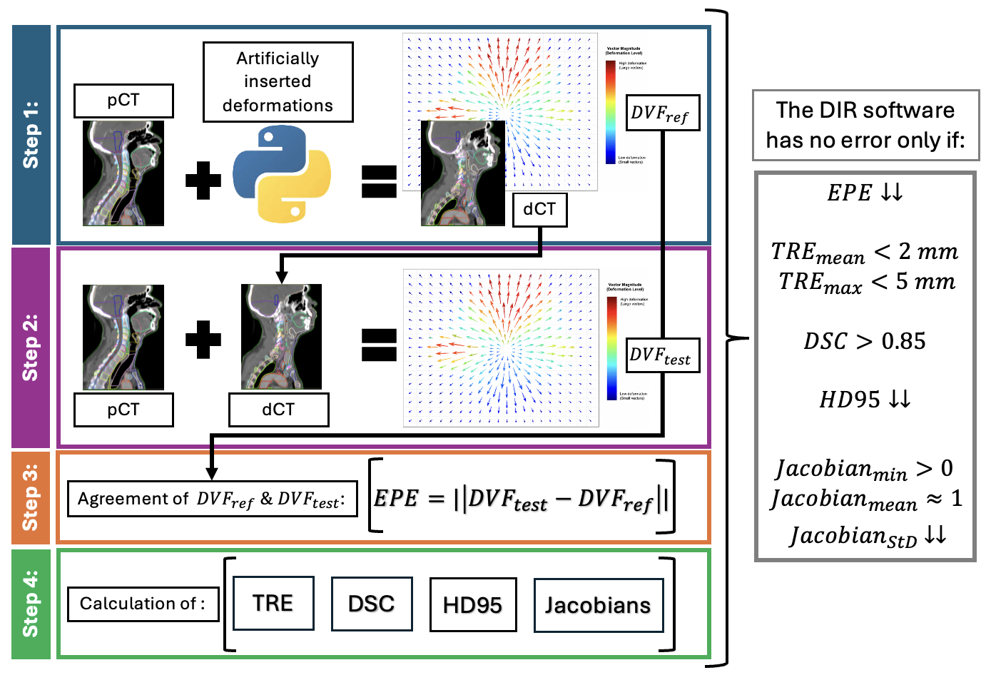
        1. Artificial Deformation Vector Field (DVF_ref) is initially
          generated using RayStation’s Organ Motion Simulation tool, then is
          defined by a user and applied to a patient’s CT (pCT) to generate
          patient-specific digital phantom, i.e. deformed CT (dCT). DVF_ref
          is considered as ground truth.
        2. The pCT and dCT are used as input to a tested DIR software to
          perform DIR and create the corresponding DVF_test.
        3. Agreement between the resulting DVF_test and DVF_ref with the help
          of End-Point Error (EPE) indicates accuracy of DIR performance,
          quantifies the registration error, and provides spatial information
          on deformation disagreement.
        4. Following the recommendations of TG-132, quantitative assessment
          is performed by automatically computing the following metrics for
          each ROI (whether autocontoured or manually delineated): Target
          Registration Error (TRE), Dice Similarity Coefficient (DSC), 95th
          percentile Hausdorff Distance (HD95), and Jacobian statistics.

        Figurem 1: Animated description of the workflow based on the combination of the two scripts on how DIR software is going to be evaluated.

        Results:

        The four-step workflow was successfully implemented. The proof of concept of the first step was visually validated in terms of correct identification of the ROIs intended for deformation and correct direction and magnitude of motion. An example of head-and-neck patient is presented in Figure 2A. Small, medium, and extreme input displacements produced corresponding deformation magnitudes, with maximum displacement consistently observed in regions adjacent to moving ROIs. Figure 2B demonstrates step-by-step validation of an individual deformation for an example of thorax patient. The validation of steps 2-4 is ongoing.
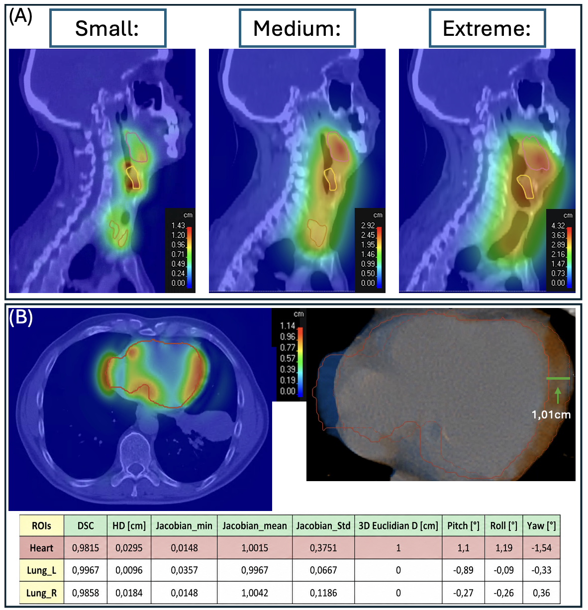
        Figure 2: (A) Example of a Head and Neck patient used as input to test the generation of digital phantom. After the autocontouring, three ROIs (Larynx, Thyroid Gland, and Tongue Base) were moved with three different magnitudes (small, medium, and extreme) to test the functionality of the script. (B) Example of detailed validation on a Thorax patient by applying an extreme displacement of 1cm only in Heart in Left-Right axis. The validation was first qualitative by visual inspection of colourwash showing the location of the deformation, followed by quantitative measurements of the distance between the reference and the moved heart structure in the fusion image. Last step was the calculation of the metrics of heart and surrounding ROIs.

        Outlook & Discussion:
        An automated comprehensive workflow for DIR testing was developed, and preliminary proof of concept validation was successful. Next steps are more comprehensive and quantitative validation, automatic recognition of anatomical regions, and identification of acceptable tolerances, mostly for EPE and the rest of the metrics. Combining patient-specific “digital-phantom” with established DIR validation metrics can potentially improve confidence in DIR clinical application and provide comprehensive description of individual’s algorithm limitations.
        Acknowledgments: CTU has a scientific collaboration with RaySearch Laboratories.

        References:
        1. Kim Haksoo, Park Samuel et al., “Quantitative Analysis Tools and Digital Phantoms for Deformable Image Registration Quality Assurance”. 2015, Technology in cancer research & treatment. 14. DOI: 10.1177/1533034614553891.
        2. Brock Kristy K., Mutic Sasa et al., “Use of image registration and fusion algorithms and techniques in radiotherapy: Report of the AAPM Radiation Therapy Committee Task Group No. 132”. 2017, Medical Physics, volume 44, issue 7. DOI: https://doi.org/10.1002/mp.12256

        Speaker: Panagiotis Alafogiannis (Dept. of Dosimetry and Appl. Of Ion. Radiation, Faculty of Nuclear Sciences and Physical Engineering, Czech Technical Univ. in Prague, Czech Republic & Dept. of Oncology, Univ. Hospital and Faculty of Medicine, Ostrava, Czech Republic)
    • 18:00
      AI4PT dinner
    • 08:15
      Joint symposium on AI4R/PT
    • 50
      Closing plenary: future of AI for RT

      Dr. Xing is currently the Jacob Haimson  & Sarah S. Donaldson Professor of Medical Physics and Director of Medical Physics Division of Radiation OncologyDepartment at Stanford University. He also holds affiliate faculty positions in Department of Electrical engineering, Institute of Computational and Mathematical Engineering (ICME), Biomedical Informatics, Human-Centered Artificial Intelligence, Bio-X and Molecular Imaging Program at Stanford. His research has been focused on medical imaging, AI in medicine, and image-guided interventions. He has made unique and significant contributions to each of the above areas. Dr. Xing is an author on more than 500 peer reviewed publications in high impact journals. He is a fellow of AAPM, ASTRO, and AIMBE. He is the recipient of the 2023 Edith Quimby Lifetime Achievement Award of AAPM.

      Speaker: Prof. Lei Xing (Stanford University)
    • Session D: Oral presentation x3
      • 51
        Real-Time Range Prediction for a Mixed Helium–Carbon Beam

        Objective

        Mixed helium-carbon beams enable online range monitoring and imaging. Both ions share the same mass-to-charge ratio, allowing simultaneous acceleration, while helium has approximately three times the range in water. However, anatomical changes beyond the carbon Bragg peak only affect the helium range. We present a deep learning approach for spot-by-spot carbon range prediction using mixed beam detector signals and planning CT information.

        Methods

        Mixed-beam simulations were performed using our custom GPU Monte Carlo code, Raymond, based on Geant4 (QGSP_BIC_HP, EM opt4). The dataset comprised two-field treatment plans on 17 patient CTs with Lung and Liver targets and anterior–posterior and posterior–anterior beam directions. Simulations used end-inhale and end-exhale (phases 00 and 05), representing the largest respiratory motion. In total, 882,350 mixed helium–carbon pencil beams with 10% helium were simulated in both phases.
        Mixed detector images were preprocessed by a separate defragmentation model to remove carbon fragment contributions. The range prediction network combined convolutional encoders and an attention layer, using the cleaned images from phases 00 and 05, beam energy, and the phase 00 CT and carbon dose profile. It predicted the phase 05 carbon dose profile for each spot, from which R80 and its change relative to phase 00 were calculated.
        Evaluation used five-fold cross-validation across patients. Mean absolute error (MAE) and root mean squared error (RMSE) were calculated per test patient and averaged separately for Lung and Liver.

        Results

        For Lung, the network achieved an MAE/RMSE of 2/4 mm, compared with 3.7/6.3 mm when assuming no carbon range change and 4.6/8.2 mm when assuming equal range changes for helium and carbon. For Liver, the network achieved 1.3/2.2 mm, compared with 2.2/3.7 mm and 2.1/3.2 mm under the respective assumptions. MAE reductions relative to these baselines were 48% and 58% for Lung, and 43% and 38% for Liver. Inference, including defragmentation and R80 calculation, took approximately 7 ms per beam on an NVIDIA H200.

        Outlook & Discussion

        Evaluation on simulated patient treatment plans demonstrated accurate carbon range prediction for both sites. Millisecond inference supports spot-by-spot assessment, highlighting the potential of mixed beams for online treatment monitoring. Future work will address clinically relevant dose changes and experimental validation.

        Speaker: Maximilian Dick (GSI Helmholtz Center for Heavy Ion Research GmbH)
      • 52
        Deep-learning assisted single-scenario robust optimization for carbon ion therapy

        Objective: Robust optimization in particle therapy conventionally uses a voxel wise-worst case approach, where the plan is optimized to meet the clinical criteria even under the worst of a set of uncertainty scenarios. This entails significant computational overhead, most of which is only needed to compute the worst-case scenario. We hypothesize that optimizing only on the nominal case and a predicted last-iteraion worst-case scenario per voxel will provide equivalent results, at significantly reduced computational load.

        Methods: 47 head&neck cancer patients were selected from the patient cohort treated in the GSI carbon ion therapy pilot project, and randomly split in training (35) and validation set (12). For each patient, full robust (FRO, 9 scenarios, 3% range and 3% setup uncertainties) and non-robust (NRO) carbon ion therapy treatment plans were optimized with the experimental treatment planning system TRiP98. For every optimization iteration, the voxel-wise worst case scenario was stored. As a proof-of-concept that single-scenario worst-case (SSRO) optimization can produce equivalent results, the scenario output of the last iteration of the FRO optimization was used as input for a robust optimization on the nominal and predetermined worst-case scenario. Robust evaluation was performed on 9 scenarios and considered typical DVH metrices. A Unet model and bespoke preprocessing workflow was developed with MONAI. The loss was a composite loss of class-weighted cross entropy and dice loss.

        Results: The robust optimization based on the last-iteration worst case scenario performed clinically equivalent to the conventional robust optimization, at mean minimum D95% near 98%, and all cases above 96%. SSRO saved about 70% of the runtime per iteration, while it reduced the memory for the dose-influence matrix by a factor 4. Conversely, non-robust optimization performed considerably worse, at mean minimum D95% of 91.7%. The deep-learning model achieved promising performance on the validation data, with strong overall spatial agreement, particularly in OARs, between the true and predicted worst case scenario. Still, predictions over-homogenize regions of considerable scenario variation in the label, leaving room for further improvement.

        Outlook & Discussion: Robust optimization based on a single scenario is a more efficient approach to robust optimization. With further fine-tuning of the deep-learning prediction, streamlined, memory efficient robust optimization becomes possible for carbon ion therapy. Future work will expand the number of robustness scenarios and test architectures tailored to locally highly variable class labels.

        Speaker: Lennart Volz (GSI Helmholtz Center for Heavy Ion Research GmbH)
      • 53
        A real association is not a usable predictor: a five-stage workflow for machine learning on routine radiotherapy QA data

        Objective
        Radiotherapy services routinely generate machine and dosimetric QA data, rarely analysed together. We propose a reproducible five-stage workflow for developing machine-learning models from such data, focusing on the step least often addressed: establishing whether exploitable signal exists, and distinguishing a genuine but weak physical association from useful predictive capability. We demonstrate it linking multileaf-collimator (MLC) technical controls with pre-treatment dosimetric QA across three linacs (727 temporally matched pairs, 2022–2025).

        Methods
        Five stages: (1) identify underused routine data; (2) integrate it preserving temporal structure; (3) model with physically interpretable features, a linear baseline and leakage-free chronological validation; (4) audit the signal rather than the error alone — explainability consistent with physics, unsupervised analysis complementing supervised models, apparent detectors tested against chance; (5) delimit validity and deployment conditions.
        Weekly MLC controls (AQUA, Elekta) were matched by linac to QA records (Verisoft, PTW). Ridge and XGBoost regressors predicted the gamma passing rate at 3%/2 mm. Audits used SHAP, per-linac principal component analysis, and a robust Mahalanobis/Hotelling T² control chart benchmarked against frequency-matched random alarms (permutation test).

        Results
        Gamma passing rates concentrated in 92–100%, leaving little exploitable variation. Ridge yielded a mean absolute error of 1.3 percentage points (pp) but R² ≈ 0.03; XGBoost performed worse than predicting the mean (R² = −0.37).
        The audits were more informative. SHAP importance was physically coherent; per-linac principal components correlated with gamma error (ρ = 0.24 and 0.20; p < 0.01). The control chart exposed the central difficulty. Flagged controls carried greater gamma error (1.4 versus 1.0 pp; p < 0.001), and Mahalanobis distance correlated with gamma error (ρ = 0.17; p < 0.001): the association was real. However, as a detector of individual episodes it did not outperform chance: depending on the lead-time window (14–60 days) it identified 5–8 of 11, matched by random alarms (p = 0.74).
        The framework nevertheless correctly characterised a real jaw-mirror malfunction (gamma = 90%), jaw variables dominating both SHAP and the chart.

        Outlook & Discussion
        The main lesson is a second-order negative result: a statistically significant but weak association can masquerade as predictive capability. In a well-maintained service, events are rare and the signal too weak for reliable individual-episode prediction.
        This yields a practical acceptance criterion: no QA prediction tool should be deployed until it outperforms an appropriate chance benchmark. Further: integrating the data required more effort than modelling them, and the workflow's demonstrated value lies in supporting diagnosis under medical-physicist supervision, not in autonomous prediction.

        Speaker: Alejandro del Pozo Domínguez
    • 10:30
      Coffee break
    • 54
      AI4RT plenary discussion
      Speakers: Prof. Aswin Hoffmann (Helmholtz-Zentrum Dresden-Rossendorf), Dr Ye Zhang (PSI - Paul Scherrer Institut)
    • 55
      Closing remark -- Lunch (to go)
    • 13:00
      Departure AI4PT (flexible)