
Speaker
Description
Objective:
Management of respiratory motion, online adaptive radiotherapy, and reirradiation require accurate and reliable Deformable Image Registrations (DIRs) to allow fast and precise structure mapping and dose accumulation. Artificial Intelligence (AI)-based DIR algorithms offer promising improvements in DIR performance. However, currently used DIR validation metrics are not sufficient to provide trustworthy validation of DIR for clinical usage. Therefore, this work aims to develop robust and automated workflow for validating DIR algorithms prior to clinical adaptation.
Methods:
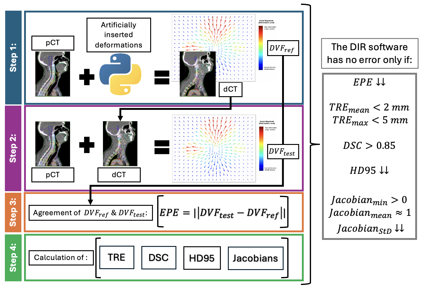
A four-step DIR validation workflow was developed using Python scripting. The schematic overview is provided in Figure 1:
- Artificial Deformation Vector Field (DVF_ref) is initially
generated using RayStation’s Organ Motion Simulation tool, then is
defined by a user and applied to a patient’s CT (pCT) to generate
patient-specific digital phantom, i.e. deformed CT (dCT). DVF_ref
is considered as ground truth. - The pCT and dCT are used as input to a tested DIR software to
perform DIR and create the corresponding DVF_test. - Agreement between the resulting DVF_test and DVF_ref with the help
of End-Point Error (EPE) indicates accuracy of DIR performance,
quantifies the registration error, and provides spatial information
on deformation disagreement. - Following the recommendations of TG-132, quantitative assessment
is performed by automatically computing the following metrics for
each ROI (whether autocontoured or manually delineated): Target
Registration Error (TRE), Dice Similarity Coefficient (DSC), 95th
percentile Hausdorff Distance (HD95), and Jacobian statistics.
Results:
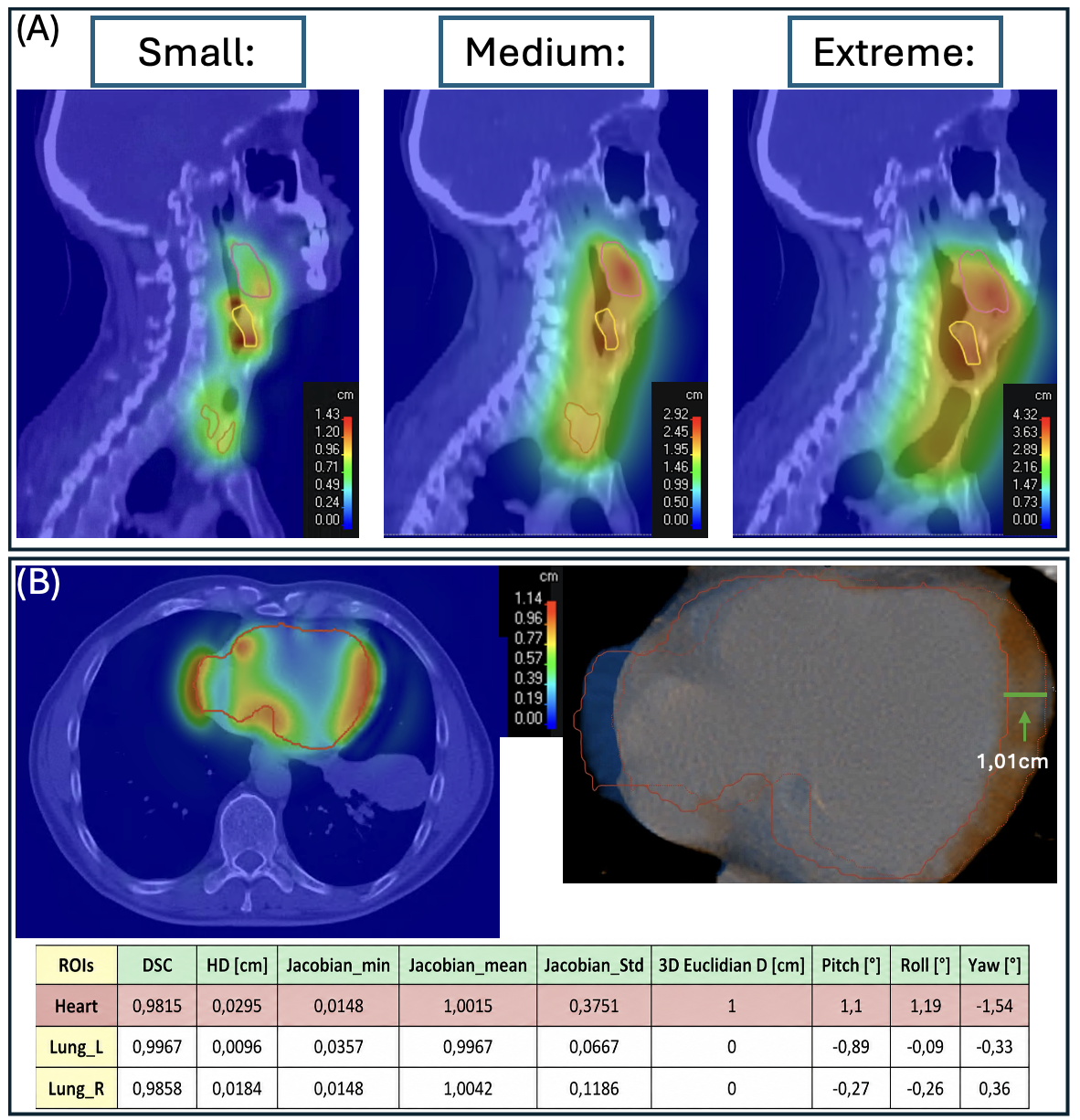
The four-step workflow was successfully implemented. The proof of concept of the first step was visually validated in terms of correct identification of the ROIs intended for deformation and correct direction and magnitude of motion. An example of head-and-neck patient is presented in Figure 2A. Small, medium, and extreme input displacements produced corresponding deformation magnitudes, with maximum displacement consistently observed in regions adjacent to moving ROIs. Figure 2B demonstrates step-by-step validation of an individual deformation for an example of thorax patient. The validation of steps 2-4 is ongoing.
Outlook & Discussion:
An automated comprehensive workflow for DIR testing was developed, and preliminary proof of concept validation was successful. Next steps are more comprehensive and quantitative validation, automatic recognition of anatomical regions, and identification of acceptable tolerances, mostly for EPE and the rest of the metrics. Combining patient-specific “digital-phantom” with established DIR validation metrics can potentially improve confidence in DIR clinical application and provide comprehensive description of individual’s algorithm limitations.
Acknowledgments: CTU has a scientific collaboration with RaySearch Laboratories.
References:
1. Kim Haksoo, Park Samuel et al., “Quantitative Analysis Tools and Digital Phantoms for Deformable Image Registration Quality Assurance”. 2015, Technology in cancer research & treatment. 14. DOI: 10.1177/1533034614553891.
2. Brock Kristy K., Mutic Sasa et al., “Use of image registration and fusion algorithms and techniques in radiotherapy: Report of the AAPM Radiation Therapy Committee Task Group No. 132”. 2017, Medical Physics, volume 44, issue 7. DOI: https://doi.org/10.1002/mp.12256