Choose timezone
Your profile timezone:
Dear Colleagues and Friends,
It is a great pleasure to invite you to the 58th Annual Meeting of the Swiss Society of Radiobiology and Medical Physics (SSRMP), held in the historic and vibrant city of St. Gallen on November 5th and 6th, 2026.
Following our society’s long-standing tradition - from our founding in Geneva in 1964 to our most recent gatherings at CERN and in Lucerne in the heart of Switzerland - we continue to evolve. Our field has seen a remarkable shift: new generations of medical physicists are stepping forward, and our focus has expanded from the fundamentals of radiation protection to cutting-edge research in patient care and interdisciplinary clinical practice.
St. Gallen, famous for its UNESCO World Heritage Abbey District and its history as a center of textile and knowledge, provides the perfect backdrop for our meeting. The city also shares a unique bond with our profession: in 1897, the first X-ray institute in Switzerland was opened here in collaboration with Wilhelm Conrad Röntgen. We aim to bridge the gap between tradition and innovation, bringing together experts in medical physics, radiobiology, and radiation protection.
Scientific Exchange & Networking An engaging scientific program is only possible through your contributions. We look forward to a diverse range of abstracts reflecting the high level of research and clinical investigation currently thriving in Switzerland.
As always, our industrial partners will accompany us throughout the meeting. We encourage you to use the breaks in our exhibition area for lively discussions, fostering the collaboration that is essential for the technical advancement of our discipline.
Cultural & Social Highlights To complement the scientific program, we have arranged a special opportunity to visit the world-famous Abbey Library (Stiftsbibliothek). The Social Event at Restaurant Seeger is located in the heart of the city - this traditional venue offers the ideal atmosphere for a relaxed evening of dinner and networking with friends and colleagues.
The SSRMP General Assembly (GA) will take place on November 5, 2026, at 16:30.
Admission to the General Assembly is open to all members free of charge and does not require conference registration.
We look forward to seeing you in St. Gallen!
The Local Organizing Committee is responsible for the overall logistics, registration, and execution of the 58th SSRMP Annual Meeting in St. Gallen.
Samuel Peters (Chair) samuel.peters@h-och.ch
Tanja Hertel
Dr. Konrad Buchauer
Simon Heinze
Liza Andrzejewska
Friedemann Herberth
The Scientific Committee is ensuring the engaging scientific program reflects the full diversity of our society.
Reto Küng, Inselspital Bern
Nikolaos Koutsouvelis, HUG
Nathan Corradini, Clinica Luganese Moncucco
Jerome Krayenbühl, USZ
Diana Wüthrich, KSGR
Veronique Valet, CHUV
Samuel Peters, HOCH Ostschweiz
Konrad Buchauer, HOCH Ostschweiz
Jennifer Griesinger, KSA/HOCH Ostschweiz
Roman Menz, USB
Saltybaeva Natalia, LUKS
Hannah Eggimann, KSA/HOCH Ostschweiz
Cester Davide, USZ
Thiago Lima, LUKS
Konstantinos Zeimpekis, Inselspital Bern
Silvano Gnesin, CHUV
Stephan Scheidegger, ZHAW Winterthur
Michaela Medowa, Bern
The SSRMP Board has officially resolved to introduce a registration fee for the upcoming conference.
| Category |
Early Bird Until |
Regular Sept 28 - Oct 28 |
Dinner Register by Oct 28 |
| SSRMP Member | 60 | 120 | 60 |
| Student | 60 | 60 | 40 |
| Non-Member | 120 | 180 | 60 |
Late/On-site Registration (from Oct 29): An additional CHF 60 will be charged on top of the regular fee.
Registration and Call for Abstracts: open
Abstract Submission Deadline: 31.8.2026 extended to 30.9.2026
Social event: 5.11.2026 19:00 Restaurant Seeger St. Gallen Das Seegerhaus in St. Gallen
Guided Tour Abbey Library (Stiftsbibliothek): 6. November 16:15 Stiftsbezirk St. Gallen - Stiftsbibliothek
Departure from the congress venue at 15:45 until 17:00
Objective: To evaluate whether GTV-PTV margin reduction facilitates biological effective dose (BED) escalation for adrenal gland metastases (AGM) stereotactic body radiotherapy (SBRT) using CBCT-guided online adaptive radiotherapy with surface-guided breath-hold (CBCT-oART-BH).
Methods: Ten AGMs were treated with CBCT-oART-BH using various fractionation schemes, 5 mm GTV-PTV margin, and +/-3 mm gating window. Simulated reference and adapted plans for the (first) 5 fractions were created for 5x8 Gy (72.00 Gy BED$_{10}$) or 8x5 Gy (62.00 Gy BED$_{10}$) prescribed to PTV-D99%, each for 5 or 3 mm GTV-PTV margin (total: 10 patients * 5 adapted plans * 4 fractionation/margin combinations). Organ-of-interest (OOI) sparing had priority over target coverage. Dosimetric endpoints and robustness against 3 mm shifts in all cardinal directions for the four fractionation/margin combinations were compared using Wilcoxon signed-rank test (5% significance level).
Results: For 5x8 Gy, acceptable PTV coverage was achieved in 3/50 adapted plans with 5 mm margin and 20/50 plans with 3 mm. For 8x5 Gy, it was achieved in 33/50 plans with 5 mm margin and 48/50 plans with 3 mm. Robustness analysis showed significantly improved GTV coverage robustness with 5 vs 3 mm margins and for 8 vs 5 fractions. Over all uncertainty scenarios, median BED$_{10}$ to GTV was significantly higher for 5x8 Gy (90.77 Gy for 5 mm margin and 89.31 Gy for 3 mm) than for 8x5 Gy (73.85 Gy for 5 mm and 72.91 Gy for 3 mm).
OOI constraint violations occurred in 5/50 adapted plans for 5x8 Gy, 3 mm; 18/50 for 5x8 Gy, 5 mm; 3/50 for 8x5 Gy, 3 mm; and 6/50 for 8x5 Gy, 5 mm. D0.03cm3 to stomach and small bowel were significantly less robust with 5 fractions than with 8; this was also true for D5cm3 to the stomach with 5 mm margin but not with 3 mm or for the small bowel with any margin.
Outlook & Discussion: Margin reduction from 5 to 3 mm enables significant BED escalation, maintaining OOI protection for AGM SBRT with CBCT-oART-BH.
Objective: Changes in daily anatomy over the course of treatment cause differences between planned and delivered dose. We propose an up-to-now (UTN) optimization offline adaptive strategy that incorporates accumulated up-to-now dose into plan adaption, compensating for inter-fraction over- or underdosage. Dynamic trajectory radiotherapy (DTRT) exploits non-coplanar beam trajectories to spare organs at risk beyond what is achievable with coplanar VMAT, while preserving target coverage. We show feasibility of UTN-optimization for offline adaptive DTRT on a clinically motivated retrospective case.
Methods: We extended the DTRT optimization process by incorporating the accumulated up-to-now dose. UTN optimization for DTRT was applied on a clinically motivated head and neck case (prescribed 50 Gy to 95% of the PTV in 25 fractions) with offline adaptive plan adaption at fraction 12. A DTRT reference plan was generated on the planning CT. Next, the dose of the reference plan was accumulated on a repeated CT at fraction 12, using an in-house dose accumulation method, and plan adaption with UTN optimization was performed. Dosimetric enpoints of the accumulated dose up to fraction 12 was compared against the total accumulated dose (the UTN dose plus the remaining 14 fractions) on the repeated CT.
Results: Accumulated dose up to fraction 12 revealed target underdosage, with a planning target volume (PTV) D95% of 97.4% and D2% of 111%. By applying UTN optimization during plan adaptation, the total accumulated dose achieved a PTV D95% of 100% and D2% of 109%. This indicates that UTN optimization successfully compensated for prior over- and underdosage. Organ-at-risk (OAR) endpoints remained similar between the delivered UTN dose and the total accumulated dose.
Outlook & Discussion: We demonstrated the feasibility of incorporating UTN optimization into offline adaptive DTRT on a first retrospective case. Integrating UTN strategies with DTRT methods shows potential to compensate for target over- and underdosage from prior fractions without compromising OAR sparing.
Supported by SNSF grant 10001696 and partly by Siemens Healthineers through its business line Varian.
Objective: Online adaptive radiotherapy (OART) is an evolving approach in prostate cancer treatment to account for daily anatomical variations. Higher doses per fraction increase the need for accurate target dose coverage and sparing of organs at risk (OARs). We retrospectively evaluated daily imaging and daily dose using automated offline dose-monitoring software to assess whether a hypothetical online dose-guided radiotherapy (DGRT) positioning strategy could improve target dose coverage compared with our conventional image-guided approach while also supporting planning target volume (PTV) margin reduction.
Methods: Sixty-nine prostate cancer patients were treated with helical tomotherapy (60 Gy in 20 fractions). PreciseART software automatically generated daily deformed contours and recalculated doses on daily kVCT or MVCT images. For each fraction, an optimal rigid translation was calculated on the daily dose volume using a two-stage grid search in-house Python algorithm that maximized the D99% of the prostate and seminal vesicle clinical target volumes (CTVs). CTV dose was evaluated at the treated and simulated DGRT positions, with D99% ≥ 95% of the prescribed dose defining clinically acceptable CTV coverage. Dose coverage for isotropic CTV expansions of 1 to 4 mm was additionally calculated to assess PTV margin reduction. Dose-volume metrics to the bladder and anorectum were compared for both approaches.
Results: In 1,345 analyzed fractions, 138 (10.3%) failed to meet the clinical acceptability in CTV dose coverage (D99% ≥ 95%) at the treated position, compared with 3 (0.2%) at the optimal DGRT position. Expanded CTV volume analysis suggested that PTV margins could be reduced by 2 mm and still maintain target coverage comparable to that achieved with the delivered treatments. OAR dose-volume metric distributions were similar for delivered and DGRT.
Discussion: The DGRT workflow improved prostate and seminal vesicle CTV dose coverage compared with conventional image-guided positioning while also suggesting potential for PTV margin reduction. In an OART setting, automated DGRT could reduce variability in user judgement during image registration and provide a more objective, dose-based positioning to decide whether plan adaptation is necessary.
Objective: Deformable image registration (DIR) is crucial in adaptive radiotherapy and dose accumulation. Quantitative validation stays challenging since the true anatomical deformation is not known a priori. We present a tool to generate desired deformed CT from reference CTs with ground-truth deformable vector fields (DVF), from single- or multi-organ deformation commands.
Methods: The tool takes the reference CT, organ contours (structure set) and the deformation command as inputs. Structures are converted into meshes, and modified following the requested deformations (translations, inflation, and shrinkage). The transformation DVF is computed through radial basis function interpolation under constraints to preserve fixed structures (e.g. bones) and prevent organs intersections and non-physiological deformations. DVF healing eliminates folding voxels in the post-processed final DVF. Finally, the reference CT and masks are warped to output: the deformed CT, corresponding deformed structure set, ground-truth DVF, Jacobian, and quality control parameters.
The tool was tested on two clinical male pelvis CTs with auto-contoured organs (Therapanacea, France). Several single- and combined- organ transformations were applied: translations up to 8 mm, inflations up to 80%, and shrinkages down to 35% for bladder, rectum and prostate. Deformed CTs were auto-contoured and the segmentations were compared with the warped original structures using Dice similarity coefficient (DSC) and 95th percentile Hausdorff distance (HD95).
Results: Deformed CTs looked realistic and achieved the desired deformations. Mean (range) DSC between auto-contoured and deformed contours were 0.971 (0.960 – 0.998) for bladder, 0.966 (0.955 – 0.978) for rectum and 0.962 (0.956 – 0.969) for prostate. Mean (range) HD95 were 2.14 mm (1.05 – 3.00) for bladder, 1.69 mm (1.05 – 3.00) for rectum and 1.11 mm (1.05 – 1.17) for prostate.
Outlook & Discussion: The tool generated anatomically realistic deformed CTs with ground-truth DVF for two male pelvis cases. The largest HD95 values corresponded to CT slice thickness and resulted from small contours on additional slices detected by auto-contouring. Other anatomical regions are still to be tested. Overall, this tool can help validate DIR and adaptive radiotherapy workflows.
Objective
To evaluate two strategies for adaptive tomohelical radiotherapy: direct dose calculation on daily kVCT using dedicated HU-to-density calibration curves, and synthetic CT (sCT) generated from daily kVCT and planning CT. Dosimetric accuracy and suitability for target and organ-at-risk (OAR) delineation were investigated.
Methods
Seventeen replanned patients (head-and-neck, n=5; thorax, n=6; pelvis, n=6) were retrospectively selected. Same-day kVCT and replanning CT (repCT) were selected, and sCT was generated in RayStation v2025 using the Corrected CBCT algorithm. Direct kVCT calculation used previously dosimetrically validated HU-to-density curves, with periodic HU stability QA (ESTRO 2026, Poster Highlight 2947). kVCT and sCT dose distributions were compared with repCT using dose metrics for 49 CTVs and 40 OARs, along with 2D gamma analysis (2%/1 mm).
For contouring assessment, nine patients (three per site) were selected. CTVs were propagated from planning CT using ANACONDA deformable image registration (DIR), while OARs were generated using DIR and deep-learning segmentation (DLS). Radiation-oncologist-corrected contours served as reference. Agreement was quantified using Dice Similarity Coefficient (DSC) and 95% Hausdorff distance (HD95).
Results
For CTVs, 50% of dose deviations relative to repCT were within ±0.5% for both strategies, with maximum deviations below 1.5%. Despite small overall deviations, kVCT-sCT differences were statistically significant (paired t-test, p<0.05) but unlikely to be clinically meaningful. No significant difference was observed for OARs (p>0.05). Direct kVCT-sCT gamma passing rates exceeded 90%, with most between 98–100%. CTV propagation was robust on both modalities, with mean DSC of 0.93–0.99 and HD95 below 1 mm. For OARs, the optimal delineation method (DIR or DLS) depended on organ type, with no clear difference between kVCT and sCT.
Outlook & Discussion
Both modalities appear suitable for adaptive radiotherapy but require different QA strategies. Direct kVCT calculation benefits from established HU-to-density calibration and periodic stability monitoring, whereas sCT introduces algorithm-related uncertainties requiring dedicated QA for each generated sCT, with appropriate procedures yet to be established. Workflow efficiency, computation time and QA burden will guide clinical implementation.
Objective
Adaptive proton therapy relies on repeated imaging to monitor anatomical changes and guide plan adaptation, making additional imaging dose important. CT enables proton dose calculation but involves ionizing radiation, whereas MRI provides superior soft-tissue contrast without radiation exposure but lacks the information required for direct proton dose calculation.
We investigated an MRI-only adaptation framework combining two recently developed components: deep-learning-based direct proton dose calculation on MRI (10.1016/j.phro.2025.100806), avoiding synthetic CT generation, and a differentiable optimizer enabling gradient-based plan re-optimization directly through the learned dose model. We experimentally assessed this combined approach under anatomical changes, using the clinical CT-based workflow as reference.
Methods
Our experimental evaluation used MRI and CT data acquired from two ex-vivo pig heads for treatment planning and algorithm fine-tuning. The direct-to-MRI dose calculation algorithm uses deep-learning to directly map MRI-derived anatomical information to Monte-Carlo-simulated proton dose. The differentiable optimizer propagates dose-objective gradients through this model to update pencil-beam parameters and fluences. For this initial validation, a single 0° beam was used, with the beam angle kept fixed. Plans delivering 2 Gy to wedge-shaped and homogeneous targets were generated.
After two days, fat layers were removed to simulate weight loss, followed by repeat MRI and CT scans. Plans were directly re-optimized on MRI using the combined framework and, for comparison, on CT following the clinical workflow.
Both plans were delivered to the pig heads with Gafchromic films for relative dosimetric measurements. Planned and measured dose distributions were compared using global gamma analysis (3%/3 mm,10% threshold).
Results
All re-optimized plans maintained target coverage, with gamma pass rates above the 95% clinical acceptance threshold: 95.5–99.0% for direct MRI-based re-optimization and 98.5–99.5% for the CT-based reference.
Outlook and Discussion
This initial validation demonstrates the feasibility of combining direct MRI-based dose calculation with differentiable plan optimization for proton adaptation after anatomical change.
By avoiding synthetic CT generation, the approach may simplify and accelerate MRI-guided adaptive proton therapy while avoiding additional imaging dose.
Further validation, dedicated quality assurance strategies and clinical workflow integration are still required.
Anatomical changes during radiotherapy treatments can compromise dose accuracy despite daily CBCT guidance. Offline adaptive radiotherapy (ART) traditionally requires time-consuming re-planning CT scans, interrupting treatment and increasing radiation exposure. Synthetic CT (sCT) generation from CBCT offers a promising alternative. This study evaluated the HU and dosimetric accuracy of three sCT methods: corrected CBCT (corrCBCT), virtual CT (vCT) integrated in RayStation 2024B, and an AI-based approach (AICT; Image+ CBCT 1.0.0). Two CBCT acquisition protocols were also assessed to determine the influence of input image quality on sCT performance.
Twenty prostate cancer patients treated with a Versa HD linac equipped with the XVI R5 imaging system (Elekta AB, Stockholm, Sweden) underwent planning CT (pCT) followed by daily CBCT guidance. Two CBCTs acquired during the first two treatment days (CTDIvol 32.2 and 16 mGy) were selected, each generating three sCTs. HU accuracy was assessed in pelvic organs and CTV against pCT. For dosimetric evaluation, clinical plans were recalculated on all sCTs and on deformed CT (dCT; ROI-constrained DIR of the pCT to the corrCBCT), used as reference. Validation included dose-volume histogram (DVH) parameters and 3D local gamma analysis (2%/2mm, 10% low-dose threshold).
All methods showed satisfactory HU agreement with pCT. Bony structures showed larger variations, while soft tissue values were highly consistent. Rectal results were more variable, reflecting anatomical variability.
Dosimetric evaluation showed clinically acceptable agreement with dCT, with DVH differences generally within ±1 Gy and ±1% and gamma pass rates ranging from 94% to 100%, except for a single rectal outlier (89.61%).
This study demonstrated that all three CBCT-based sCT generation methods substantially improved HU accuracy and achieved clinically acceptable dose calculations for ART. No clinically relevant differences were observed between the evaluated methods or between the two CBCT acquisition protocols, indicating that lower-dose CBCT images remain suitable for sCT generation.
Among the investigated approaches, the AI-based method achieved comparable dosimetric performance while operating independently of the pCT and providing automatic segmentation, which may facilitate its integration into offline and online ART workflows.
Objective: To implement an independent Monte Carlo (MC), dose warping based dose accumulation framework for online adaptive radiotherapy, evaluate its robustness against deformable image registration (DIR) uncertainties, and compare it against the clinically accumulated dose from ETHOS.
Methods: The framework was applied retrospectively to two esophageal cancer cases (50.4 Gy to PTV D50%, 28 fractions). For this purpose, the dose of each online adapted plan was recalculated with the Swiss Monte Carlo Plan (SMCP) on the synthetic CT of the daily CBCT. The daily sCT was deformably registered to the planning CT and the resulting deformation vector field was used to warp each daily dose onto the planning CT for accumulation. Robustness against DIR uncertainty was assessed by repeating registration and accumulation across six DIR configurations varying regularization and optimization parameters, while evaluating the resulting variation in dosimetric endpoints. In addition, dosimetric endpoints were compared between the dose accumulated with the novel framework and ETHOS. To assess whether the registration was plausible, Dice similarity coefficient (DSC) and Hausdorff distance were computed.
Results: Across the six DIR configurations, accumulated PTV D50% varied by less than 0.1% in both cases. The largest dose differences between the six DIR configurations occurred in structures in or near steep dose gradients, where small registration differences translate into large dose differences. Differences to ETHOS in mean dose to the heart, liver, and kidneys generally exceeded the corresponding standard deviation across DIR configurations except for the kidneys in one case, where these differences were similar. Compared with ETHOS, the warped dose showed worse PTV coverage in both cases. DSC and Hausdorff distance were of limited use for evaluating DIR quality, as both metrics were strongly affected by contour inconsistencies.
Outlook & Discussion: An independent MC, dose warping based accumulation framework was developed and applied for two cases, showing that the chosen DIR configuration had only a minor influence on the accumulated dose.
Supported by SNSF grant 10001696 and partly by Siemens Healthineers through its business line Varian.
Objective: Temporally feathered radiation therapy (TFRT) is an emerging treatment concept in which multiple iso-curative subplans are used over a treatment course to intentionally modulate fractional doses to healthy organs. Dynamic normal tissue complication probability (dNTCP) modelling predicts that high-to-low interfractional dose modulation enhances normal-tissue recovery, reducing toxicity relative to established techniques. This study introduces a TFRT planning concept for non-small cell lung cancer (NSCLC).
Methods: A reference conventional VMAT plan was created for a locally advanced, right-sided NSCLC case prescribing 32 x 2 Gy to the median planning target volume (PTV). Two TFRT courses were created attempting to reduce the probability of pneumonitis and esophagitis: TFRT-LR feathers the left lung (LL) and right lung (RL) in two subplans, and TFRT-LRE feathers the oesophagus in an additional third subplan. Subplan-specific optimisation objectives were derived from the reference plan objectives aiming for high-to-low dose modulation of 10% of the prescribed dose. Objectives from the reference plan were used to control the cumulative dose of the total plan. Feathering performance was quantified by the feathered organ mean dose (Dmean) differences between high and low dose fractions.
Results: All TFRT subplans were iso-curative with target D98% within 0.5% of the reference plan. TFRT-LR achieved a high-to-low Dmean difference of 3.6% of the prescription dose in LL and 1.6% in RL. TFRT-LRE achieved median Dmean differences across subplans of 1.2% for LL, 0.9% for RL and 1.7% for oesophagus. Compared with the reference plan, cumulative Dmean was reduced by 0.4% on average in the LL and RL and by 1.9% in the oesophagus. PTV homogeneity indices (ICRU83) were below 0.063 in both TFRT courses compared to 0.066 in the reference plan.
Outlook & Discussion: Iso-curative TFRT subplans with lung and oesophageal dose modulation were successfully generated. The dNTCP is expected to reduce as the dose modulation increases. Future dNTCP analysis will be used to quantify the predicted biological benefit of the modulated schedules.
Supported by the Swiss Cancer Research Foundation (KFS-6163-08-2024).
As part of the commissioning of cone-based functional radiosurgery, we evaluated the geometric accuracy of surface-guided radiation therapy for intrafraction monitoring and couch-angle-dependent position correction. SGRT accuracy was assessed using controlled translations of a head phantom, while the effectiveness of SGRT-guided corrections was independently evaluated using cone-based Winston–Lutz performed with the Steev phantom. The SGRT system detected phantom displacements with residual errors of approximately 0.1 mm. Applying SGRT-derived corrections after couch rotation reduced the measured isocentre excursion from 0.5 mm to 0.2 mm in diameter. These results support the use of SGRT as a geometrically accurate monitoring and correction solution for cone-based functional radiosurgery.
Objective The T2 dose-rate and gantry-speed (DRGS) test is widely used for VMAT linac quality assurance. However, systematic differences between inner and outer test bands are commonly observed and may reflect limitations of the test methodology rather than true delivery errors. This study investigated the origin of these deviations and evaluated modifications intended to improve the specificity and interpretability of the DRGS test.
Methods DRGS plans were delivered on three Varian TrueBeam Edge linacs equipped with HDMLC and measured using an EPID. A fluence correction was developed to compensate for reduced effective leaf travel at the field boundaries. In addition, a static-gantry reference field with identical MLC control points was evaluated as an alternative normalization strategy. Results from the original methodology, fluence-corrected analysis, and static-field normalization were compared using band-wise deviations and peak-to-peak variation.
Results The original methodology exhibited peak-to-peak variations of 1.03–1.20% across the three linacs. Following fluence correction, the variation decreased to 0.41–0.63%, indicating that a substantial proportion of the observed response originated from systematic edge-band bias. Static-field normalization further reduced residual variation to 0.21–0.45%, suggesting additional contributions from transmission and scatter effects associated with the reference field. Absolute comparison between dynamic and static deliveries revealed small global offsets (−0.15% to +0.12%) that were not captured by conventional mean-referenced analysis. All observed effects substantially exceeded measurement repeatability (band standard deviation ≈ 0.01%).
Outlook & Discussion The results demonstrate that the original DRGS methodology is influenced by systematic effects arising from the test design and analysis approach. The proposed fluence correction substantially reduces these effects, while alternative normalization strategies provide improved separation between modulation-dependent delivery performance and other dosimetric contributors. These findings improve the interpretability of the DRGS test and may support future refinement of VMAT dynamic delivery QA procedures.
Background and purpose: Anatomical changes during head and neck radiotherapy may cause differences between planned and delivered salivary gland doses. Dose accumulation can account for these changes, but its clinical value for dose–toxicity modelling remains unclear. We investigated whether dose accumulation approaches strengthen the association of parotid dose with xerostomia compared with planned dose.
Materials and methods: Prospective data from 41 patients treated with weekly magnetic resonance guided adaptive radiotherapy within the MARTHA trial were analysed. Parotid mean doses were calculated using different reference anatomies, image registrations, and dose summation methods. The primary endpoint was Grade ≥2 clinician-rated xerostomia at 12 months, with patient-reported xerostomia as a secondary endpoint. Xerostomia rates were measured using the LENT-SOMA side-effect scale. For each strategy, ipsilateral and contralateral parotid doses were included simultaneously in binary logistic regression models. Discrimination was assessed using the area under the receiver operating characteristic curve with bootstrap-derived 95% confidence intervals.
Results: Parotid mean doses ranged from 26.8–28.1 Gy ipsilaterally and 17.3–18.1 Gy contralaterally, without significant differences between strategies (P=0.084 and P=0.224, respectively). Ipsilateral dose showed stronger associations with clinician-rated xerostomia than contralateral dose. Model discrimination was similar across strategies (AUC, 0.71–0.75), with no significant improvement over planned dose. No significant dose–toxicity associations were observed for patient-reported xerostomia.
Conclusion: Adaptive dose accumulation strategies did not significantly alter parotid dose estimates or improve xerostomia prediction compared with planned dose. Within this cohort, dose–toxicity modelling was relatively insensitive to accumulation methodology, while differing associations between clinician-rated and patient-reported xerostomia highlighted the importance of endpoint selection.
Objectives : In our department, SRS multi targets are commonly treated using different isocenters. Multi targets treated in one isocenter remained more challenging due to variable size of target and distance to isocentre. PSQA protocols for SRS plans include two different detectors: Electronic Portal Imaging Device (EPID) and Octavius 4D 1600 SRS. The aim of this work was to evaluate their sensitivity and the ability of PSQA protocols to detect deliberately introduced errors and evaluate our analysis criteria for multi targets treated in one isocenter.
Methods : SEAFARER multimet brain study was used for a 10FFF beam used clinically on a Truebeam EDGE accelerator equipped with a HD MLC. This study included three targets treated in a single isocenter (6mm, 10mm, 17mm diameters). Original plan was a clinical plan and modified plans were derived from original plan with introduced errors on MLC, MU or collimator, gantry, couch rotations. A change in DVH parameters > 5% was used as a threshold to determine a “should fail” plan. Portal Dosimetry Image Prediction 16.1 and Verisoft 8.1 were used for analysis. An in-house table was used to correct Octavius 1600 SRS response for field size and dose rate. Evaluation was performed with global gamma index using a 3%/1mm and 5%/1mm criterion, with a 10% dose threshold. Acceptance rate was gamma ≥ 95% (“pass” plan).
Results : 6 plans out of 21 highlighted at least one protocol not able to detect introduced error. 3 “should fail” plans were validated as “pass” with both protocols. Main limitations were observed for MLC errors ≤ 0.5mm and gantry, collimator angle errors ≤ 1°. Retrospective analysis with optimised criteria for Octavius SRS measurements resulted in one plan unable to detect a 1° gantry error.
Discussion : Evaluating PSQA protocols limitations seemed crucial. This work was used to fine-tune our analysis criteria for multi targets of variable diameter treated in a single isocenter. Combining two different PSQA methods ensured robustness in better identifying potential errors on clinical plans.
Objective:
Photon-counting computed tomography (PCCT) combines improved detector efficiency, spectral imaging capabilities, and ultra-high-resolution (UHR) acquisition, offering new opportunities for optimizing low-dose thoracic CT protocols. In parallel, spectral shaping using tin (Sn) filtration has been introduced to improve dose efficiency in high-contrast examinations. This study evaluated the impact of acquisition mode, reconstruction strategy, and tin filtration on image quality and lesion detectability in low-dose non-contrast chest CT using a phantom-based PCCT workflow.
Methods:
Experiments were performed on a NAEOTOM Alpha.Pro photon-counting CT system. Low-dose acquisitions were performed using four clinically available acquisition modes: Q+120, Q+140, QSn100, and QSn140. Three phantoms (Catphan® 504, Lungman, and QRM Spectral CT Phantom II) were scanned and reconstructed using regular-resolution and ultra-high-resolution (UHR) acquisition modes. Full-spectrum (T3D) reconstructions were compared with monoenergetic reconstructions at 73 keV. Image quality was characterized using the noise power spectrum (NPS), contrast-to-noise ratio (CNR), task transfer function (TTF), and line-pair analysis. Lesion detectability was assessed using a non-prewhitening model observer, and diagnostic performance was quantified using the area under the receiver operating characteristic curve (AUC). Blinded subjective evaluatioon was performed by 3 radiologists, 2 radiographers and 2 pulmonologists using the lungman phantom.
Results:
UHR acquisition consistently improved image quality compared with regular-resolution imaging. Noise was reduced, while CNR, spatial resolution, and lesion detectability increased, particularly for low-density and high-contrast structures such as lung tissue. These improvements translated into higher AUC values, especially for small lesions. Full-spectrum (T3D) reconstructions showed slightly better detectability and AUC than monoenergetic reconstructions, with the largest advantage observed for iodinated inserts.
Tin filtration had minimal impact on noise magnitude and texture but generally reduced CNR, detectability, and AUC for soft-tissue and iodinated inserts. In contrast, lung-equivalent material showed improved performance with tin filtration. Among all evaluated combinations, Q+140 reconstructed in UHR mode yielded the highest overall AUC values, demonstrating the best balance between contrast, spatial resolution, and lesion detectability.
Outlook & Discussion:
These results suggest that UHR acquisition is the primary contributor to performance gains in low-dose PCCT chest imaging. While tin filtration remains beneficial for selected high-contrast applications, Q+140 combined with UHR acquisition provided the best overall diagnostic performance. The phantom-based findings were further validated through a blinded image quality assessment involving three radiologists, two radiographers, and two pulmonologists. The subjective evaluation was consistent with the objective image quality metrics and confirmed the superior overall performance of the Q+140 UHR protocol. Larger clinical studies are warranted to confirm these results in patient imaging.
Objective
A gantry-based, full-body CBCT for diagnostic imaging has recently been introduced; our institute is assessing its clinical potential compared to CT.
Methods
Three different anthropomorphic phantoms (hand with bones, torso with full spine and chest with lungs and noduli) were imaged with different manual protocols of CBCT, the clinical protocol of a conventional CT (EICT) and the clinical protocol of a photon-counting CT (PCCT). The images were compared in terms of dose, visual assessment and objective measurements (noise, nodule volumetry).
Results
With the hand phantom CBCT showed the lowest dose values (0.37 CTDIvol), followed by EICT and PCCT (0.76 and 3.56 mGy, respectively). Absolute noise was lowest for PCCT (15.1), followed by CBCT (23.2). Qualitatively, CBCT generally achieved the best scores.
Regarding the spine CBCT achieved diagnostic-quality imaging. Subjective image quality was highest for PCCT, followed by EICT and CBCT. CBCT demonstrated lower noise than EICT, nearing PCCT levels. High radiation doses (5 mGy) were required for CBCT imaging of the upper thoracic spine due to anatomical complexity, while low doses (0,5 mGy) sufficed for the lower thoracolumbar spine.
In the lung noduli scenario, CBCT diagnostic acceptability was achieved between 98.8% and 81.0% of ratings depending on the dose level. Median absolute percentage error for noduli volumetry ranged from 8.9% to 20.3% for CBCT compared with 4.0% to 5.1% for PCCT. Radiation dose ranged from 0.40 to 4.06 mGy for CBCT, compared with 0.50 mGy for ultra-low-dose and 0.85 mGy for low-dose PCCT.
Outlook & Discussion
For extremities this CBCT can provide detailed depiction of fine osseous structures, demonstrating comparable or lower doses compared to CT.
CBCT was able to generate diagnostic-quality images of large spinal regions at low radiation doses, although the upper thoracic spine required higher doses. The overall subjective image quality remained below EICT and PCCT.
Whole-lung phantom imaging with CBCT was technically feasible and maintained diagnostic visibility of pulmonary nodules across several acquisition settings. PCCT provided superior image quality and more accurate and consistent volumetric measurements.
Objective : Anatomically-realistic phantoms are potential candidates for including anatomical texture in optimizations of dose and image quality in CT. Our objective is to assess the impact of using such a phantom on these optimizations, instead of a non-textured phantom.
Methods : Two inkjet-printed phantoms, one without texture and one with texture, both with low-contrast lesions of 12 and 8 mm in diameter, were CT scanned multiple times and reconstructed with a linear, an iterative and a deep-learning algorithm. These acquisitions provided 40-pixel regions-of-interest (ROIs) given to a channelized Hotelling observer (CHO) shown to predict human performance in small anatomical liver ROIs. The performance of the CHO in each phantom served as the basis for optimizing doses on a per lesion-size-and-contrast basis. Two optimization scenarios reduced dose from an arbitrary starting dose (7.1 mGy): the first until a fraction of the phantom's image quality at the starting dose was attained, the second until a fixed image quality threshold was attained in both phantoms. Finally, CHO performance and optimized doses were compared across both phantoms.
Results : CHO performance depended significantly on dose, lesion contrast and size. CHO performance for 12 mm lesions was significantly greater with the non-textured phantom than with the textured phantom, by 15 to 20% depending on the reconstruction algorithm. Optimized doses in the first scenario were not significantly different across phantoms, but in the second the optimized doses differed by 0.5 to 1.9 mGy. Relative to the optimized dose of the textured phantom, these differences ranged from 11% to 41%.
Outlook & Discussion : The optimization method and the phantom used impact the optimized doses and image quality. Provided that the textured phantom gives an estimate of image quality in patients, estimating a dose reduction with the non-textured phantom could lead to an undesired loss of detection performance in the clinic. Such a loss could be reduced by using realistically textured phantoms, thereby further helping to bridge medical physicists’ image quality evaluations with radiologists’ clinical realities.
Objective
To optimize the protocol settings of three CT coronary angiography (CTCA) acquisition techniques and to optimize acquisition technique selection for individual patients.
Methods
All CTCA exams were performed on a dual-source 2x192-slice CT scanner (SOMATOM Force, Siemens Healthineers), using three different acquisition techniques: low-pitch spiral scans with electrocardiogram (ECG)-based tube current modulation (‘Spiral’), prospectively ECG-triggered sequential step-and-shoot scans (‘Sequential’) and high-pitch ECG-triggered single heartbeat spiral scans (‘Flash’).
The three acquisition techniques were mainly optimized by using lower tube voltages set by automatic tube voltage selection instead of manual BMI-based tube voltage selection. Additionally, tube current settings were optimized.
Next, acquisition technique assignment for individual patients was optimized. Spiral and Sequential acquisitions were almost completely replaced by two Flash acquisitions (diastolic and systolic), also for high and irregular heart rates.
Optimizations were performed between August 2021 and October 2025 and monitored by an interdisciplinary team using dose management software (Radimetrics, Bayer) and clinical image quality evaluation. Effective dose (ED) estimates were calculated using Radimetrics.
Results
We analyzed 429 exams before and 758 exams after optimization. Radiation dose was substantially reduced for each acquisition technique: The mean dose length product (DLP) decreased from 72.6 mGy·cm to 47.7 mGy·cm (-34%) for Flash scans, from 287 mGy·cm to 170 mGy·cm (-41%) for Sequential scans and from 784 mGy·cm to 650 mGy·cm (-17%) for Spiral scans.
With optimized individual acquisition techniques and optimized selection of these acquisition techniques, the mean DLP (mean ED) for a complete CTCA exam (typically including a localizer radiograph, a calcium scoring acquisition, bolus tracking acquisitions and one or more CTCA acquisitions) decreased by 62 % (65 %) from 228 mGy·cm to 87 mGy·cm (4.9 mSv to 1.7 mSv).
Outlook & Discussion
Radiation doses of individual CTCA acquisitions and of complete CTCA exams were substantially reduced. The complexity of the examination workflows was reduced. In future work, optimization across additional acquisition techniques on other CT scanners by various manufacturers could further improve radiation doses and diagnostic information obtained with CTCA.
Background and aims
The O-armTM (Medtronic) is an intraoperative cone-beam CT system widely used for spinal and neurosurgical procedures. A recent software update (version 4.3) introduces two new features: Spine Smart Dose (SSD), which reduces radiation dose during 3D acquisitions by decreasing the number of projections, and 3D Long Scan (3DLS), enabling extended longitudinal coverage in a single acquisition. This study independently evaluated the impact of these new features on radiation dose and image quality before clinical implementation.
Methods and materials
Measurements were performed on the new O-armTM system using dedicated phantoms. Dose-length product was measured in air at the isocenter using a pencil ionization chamber for thorax and pelvis protocols using the available patient size setting (S, M, L, XL). Standard acquisition modes (low-dose, standard, HD) were compared with SSD mode. Image quality was assessed using a Catphan phantom through homogeneity measurements and task-transfer function analysis of spatial resolution and contrast for bone, air and soft tissue. Staff dose rates were also measured during 3D acquisitions at 1m and behind the gantry. For 3DLS, overlap between consecutive irradiated volumes and the associated additional dose were quantified.
Results
SSD mode reduced dose by 46%, 74% and 83% compared with low-dose, standard and HD modes respectively. A corresponding reduction in staff dose was observed. Image quality remained comparable to low-dose mode. During 3DLS acquisitions, dose increased by a factor of 1.6 within a 3 cm overlap region.
New O-armTM features enable significant dose reduction while maintaining adequate image quality for surgery guidance.
Keywords: O-arm; Image quality; Radiation dose reduction; Cone-beam CT; occupational exposure
Objective:
To investigate an artefact observed in HyperSight CBCT imaging on a TrueBeam linear accelerator, initially suspected to originate from an unknown high-density implant within the patient. The artefact represents itself as bright circles overlaid on the patient anatomy in all axial slices with changing diameter in head-feet direction. This work demonstrates that the artefact is instead associated with pre-scanning kV imaging. A phantom-based method for reproduction and prevention of the artefact is presented.
Methods:
An Alderson phantom was positioned at the machine isocenter. To analyse the impact of prior imaging on the artefacts, different acquisition protocols were compared:
• Half-fan CBCT scan without any prior kV-image acquisition as baseline
• Half-fan CBCT scan with a lateral kV-image prior to the scan, exposing large parts of the kV-detector to the unattenuated beam.
• Half-fan CBCT scan with an anterior kV-image prior to the CBCT acquisition. Exposed detector parts seeing the unattenuated beam were smaller compared to the kV-lateral protocol.
Results:
Artefacts were exclusively observed when a half-fan CBCT scan was preceded by lateral kV imaging. For the CBCT scan, the kV-detector shifts so that the previously highly irradiated region is close to the centre of rotation.
In the sagittal view, these artefacts appeared parallel to the phantom’s contour and were highly pronounced in the central scan region. Additionally, the longitudinal diameter of the artefacts exhibited distinct spatial variations. No such artefacts were visible using the baseline protocol and with anterior kV preparation.
Outlook & Discussion:
The observed artefacts seem related to the large difference in exposure between attenuated and unattenuated regions during pre-CBCT imaging. To avoid these artefacts, limiting the region of interest in kV image acquisition is recommended so beam exposure occurs only within the patient. Alternatively, AP kV-imaging should be preferred for pre-treatment verification, as it distributes the radiation load more uniformly.
Given that HyperSight CBCT utilizes a novel detector system, future investigations should evaluate whether this problem also occurs with conventional, older flat-panel detectors in clinical settings.
Objective: The introduction of HyperSight cone-beam CT (CBCT) on the Varian TrueBeam v4.1 platform provides improved image quality and Hounsfield unit (HU) accuracy. This study evaluates the suitability of HyperSight CBCT for direct dose calculation in locoregional breast radiotherapy, focusing on HU-to-relative electron density (RED) calibration, stability, image quality, and preliminary clinical feasibility.
Methods: A two-step evaluation was performed. First, a phantom-based commissioning study used a CIRS 062M phantom to assess the HU-to-RED relationship, with solid-water slabs added to reproduce scatter conditions. Image quality, spatial resolution, and contrast-to-noise ratio (CNR) were evaluated using a Catphan 504 phantom. Second, a clinical assessment of direct dose calculation using HyperSight CBCT is ongoing. In the first clinical cases evaluated, radiation therapy technologists visually assessed image quality. Target volumes and organs at risk (OARs) were propagated using deformable image registration, and dosimetric parameters were compared with the original planning CT (pCT).
Results: Phantom measurements demonstrated a stable HU response across different mAs settings, with a mean tissue attenuation deviation of (−0.3 ± 1.0) %. The Catphan 504 assessment showed a Modulation Transfer Function (MTF)10% of 0.37 lp/mm. Low-contrast detectability improved with higher mAs, although CNR remained low at a 1mm voxel size. Preliminary clinical evaluation confirmed the feasibility of direct dose calculation, demonstrating good agreement with clinical goals for targets and OARs. Observed discrepancies were mainly associated with anatomical changes, arm-position variations, or peripheral CBCT truncation.
Outlook & Discussion: To the best of our knowledge, this study is among the first to investigate the use of TrueBeam HyperSight CBCT for direct dose calculation in locoregional breast radiotherapy. HyperSight CBCT provides a stable HU-to-RED relationship and suitable image quality for direct dose calculation. Further development of integrated adaptive workflows, mitigation of truncation artefacts, and validation in larger patient cohorts will be essential before widespread clinical adoption.
Objective
To assess different approaches for plan-specific QA (PSQA) and compare these to the radiotherapy community.
Methods
The sensitivity and specificity of three PSQA methods was investigated within the SEAFARER audit [1]. Modified copies of a reference plan introducing delivery errors that affected the planned dose were verified for eleven VMAT head and neck (HN) and twenty two vertebral spine stereotactic (SABR) plans with Varian Portal Dosimetry (PD), SunNuclear perFraction (F#0) and PTW Octavius4D 1600-SRS (O4D). Gamma analysis was carried out using variable criteria, global normalisation and 10% low dose threshold.
Changes greater than 3% in D95%,PTV, D90%,PTV or D0.03cc,OAR determined whether a plan was a "should pass" or "should fail". This way, sensitivity and specificity of the different PSQA methodologies was assessed.
Results
For HN plans, PSQA (3% / 2mm) with F#0, PD and O4D had a sensitivity of 88%, 63% and 100% respectively, The only "should fail" plan passing the PSQA with F#0 was a plan with a 3.7% decrease in D95%,PTV as a result of 1mm change in MLC bank position. This plan was a PSQA pass for 58% of the community.
For SABR plans, PSQA (5% / 1mm) with F#0, PD and O4D had a sensitivity of 92%, 75% and 58% respectively. The only "should fail" plan passing the PSQA with F#0 was a plan with 7.2% increase in D0.03cc,spinal cord as a result of a 0.5 mm change in MLC leaf bank position. This was a PSQA pass for 55% of the community.
Specificity was 100% in all cases.
Summary & conclusion
The sensitivity (~90%) and specificity (100%) of PSQA with F#0 for VMAT plans (gamma criteria 3% / 2mm) and SABR plans (gamma criteria 5% / 1mm) agreed with at least 50% of the community. PSQA with PD or O4D was less sensitive in catching errors in delivery.
References
1. Lehman J., et. Al., Rad. Onc. 171 (2022), 121-128, https://doi.org/10.1016/j.radonc.2022.04.019
Objective: Process-driven Failure-Modes-and-Effect-Analysis (pFMEA) can prioritize hazards, but conventional implementations do not provide a common reference for comparing alternative QA workflows. We developed a baseline-anchored, phase-specific formalism that asks: how does each patient-specific quality assurance (PSQA) strategy reshape the same pencil-beam-scanning proton therapy (PBS-PT) risk landscape, including risks altered by the QA workflow itself?
Methods: A generic workflow of 20 PSQA-influenced process steps comprising 44 associated FMs was expert-scored for occurrence, severity, and detectability under NoQA and three PSQA strategies: measurement-based (Meas), log-file-based (Log), and independent secondary dose calculation (ISDC). The formalism separates preparatory Data-phase perturbations, incremental Full-phase verification, and the cumulative (Cum) NoQA-to-Full endpoint. The Data-to-Cum bridge, obtained by subtracting the Data-phase effect from Cum, expresses the Full-phase verification contribution on the common NoQA scale. FM effects were propagated to process-step, workflow-region, and full-workflow metrics using phase-consistent baseline-burden weights. Each relative aggregate equals the corresponding absolute risk change divided by baseline burden, enabling algebraic auditability and preventing low-burden FMs from dominating through percentages alone. Only the cumulative endpoint supports strict inter-method comparison; Full-phase effects remain method-specific.
Results: The framework revealed what a single final RPN comparison hides. Meas introduced the largest global Data burden (+4.5%) but the strongest bridge mitigation (-39.2%), yielding a cumulative risk-reduction of -34.7%. Log combined low Data friction (-2.9%) with a strong bridge (-37.4%), producing the largest cumulative risk-mitigation (-40.3%). ISDC was selective (+1.6%, -10.1%, and -8.5%, respectively), with greatest benefit in image- and calculation-sensitive steps. Local decomposition showed that global values arise from distinct workflow-region signatures rather than interchangeable test performance.
Outlook & Discussion: The formalism extends pFMEA from hazard prioritizations to a traceable comparison of risk-control architectures. It is rigorous but semi-quantitative: results are effect-size indices, not calibrated probabilities or a universal replacement hierarchy. Its value lies in reproducible trade-offs, a hybrid QA design, computer-aided implementation, and feedback-updated models. Requiring only a common baseline, staged interventions, and nested risk units, it may generalize beyond PT and EBRT to other complex, safety-critical socio-technical systems.
Objective:
Artificial intelligence (AI) is increasingly integrated into medical physics (MP) workflows, but its current use, users’ knowledge, and perceptions within the Swiss MP community remain poorly characterized. This study aimed to establish a national baseline of AI adoption, self-assessed knowledge, confidence, perceived benefits and barriers, and educational needs among medical physicists (MPs) in Switzerland.
Methods:
A 20-item anonymous online survey was developed by the SSRMP AI Working Group and distributed to SSRMP members between October 2025 and February 2026. Questions addressed respondent characteristics, AI knowledge and training, current clinical use, frequency of interaction, confidence in AI outputs, perceived benefits and barriers, attitudes towards AI, adoption intent, and educational priorities. Associations between variables were explored using Spearman’s rank correlation coefficient.
Results:
Seventy-four MPs completed the survey. AI was used in the department by 81% of respondents. Image segmentation/contouring was the most frequently reported application (78%), followed by treatment planning (31%), image registration (19%), and adaptive radiotherapy (16%). Self-assessed AI literacy was moderate, with a mean literacy index of 50±23, and only 42% reported formal AI training. AI literacy was positively associated with confidence in AI outputs (ρ=0.33, p=0.004) and five-year adoption intent (ρ=0.34, p=0.003). AI use was more frequent among respondents involved in radiotherapy (ρ=0.44, p<0.001), while years of professional experience showed no significant association with AI use or literacy. Main perceived benefits were time and workload reduction, automated segmentation, and improved consistency and reproducibility. Principal barriers were limited interpretability, lack of clinical validation/generalizability, and insufficient training or expertise. Standardized validation frameworks and clinical benchmarking were identified as the missing developments, while clinical implementation and validation was the most requested educational topic.
Outlook:
This first national survey provides a baseline of AI adoption and perceptions among Swiss MPs. Findings highlight widespread clinical exposure to AI alongside needs for education, validation frameworks, and clinical guidance. These results can support SSRMP educational activities and provide a reference for longitudinal assessment of AI adoption and competencies within the Swiss MP community.
Objective
Deep learning promises to accelerate radiotherapy planning, yet the literature conflates two distinct tasks: dose prediction, which estimates an achievable dose from anatomy alone and has many clinically acceptable solutions, and dose calculation, which computes the dose of a fully specified plan with a single physically determined ground truth. Two model families dominate. U-Nets map the input to one dose distribution in a single forward pass, whereas diffusion models (DDPMs) generate dose by iteratively denoising random noise conditioned on the patient, requiring many network evaluations. We compare both model families on both tasks (dose prediction and calculation) under identical conditions.
Methods
Five U-Nets and five DDPMs were trained and evaluated identically on 472 head and neck VMAT plans from the GDP-HMM dataset. Inputs were CT and target and OAR structures, with beam geometry added for dose calculation. To evaluate these 10 models, we developed a task-specific evaluation framework of up to eight clinically grounded metrics per task. These metrics were defined together with medical physicists and cover dose-volume histograms, OAR constraints, conformity, dose gradient, homogeneity and voxel-wise comparison.
Results
Neither model family dominates: differences within model families exceed those between them. The established U-Net DeepLabv3+ is statistically indistinguishable from the best DDPM on several metrics at a thousandth of the sampling cost. Among DDPMs, BeamDiff leads dose prediction and SP-DiffDose the target-focused dose calculation metrics. No single metric suffices, as constraint satisfaction and homogeneity rank patients and models almost independently of the others. All models share the same failure modes: overpredicted OAR dose, hot spots absent from the reference plan, and missing beam-induced structure. Models largely fail on the same volumes across tasks.
Outlook & Discussion
Shared failure modes implicate the pixel-wise training objective rather than the model, and shared failing volumes indicate that difficulty lies in the data. Model family is therefore not the decisive choice, and a single global score hides clinically relevant differences. Future work should explore training objectives beyond pixel-wise losses and faster diffusion sampling.
Objective
A novel C-arm linac was introduced. The Varian Accela linac on one hand improves hardware with a higher maximum gantry speed (15 deg/s), dose rate (40 Gy/min), MLC speed in double-stack (6.25 cm/s) improved imaging (HyperSight) and conical collimators re-design; and on the other hand introduces software integration for increased efficiency in treatment planning and adaption, patient positioning, quality assurance and commissioning (golden beam data). While these features are promising, they require a prospective evaluation. We report on linac and beam model commissioning.
Methods
The Accela SN1008 has been investigated in pre-clinical and clinical commissioning since April 2026. The output was calibrated according to SSRMP Rec. 8 with the TRS-483 extension for non-standard fields (10.5x10.5 cm2) and verified externally (IROC). Beam profiles, PDD and output factors were acquired for MLC fields ranging from 40x40 cm2 down to 0.5x0.75 cm2 ; for cones of diameter 10, 7.5, 5 and 4 mm and compared to the TPS. DLG was measured for the proximal, distal and combined MLC.
Results
The output calibration was confirmed within 2% for all beam qualities (OSLD uncertainty=3.2%, k=2). PDD and profiles for MLC fields between 40x40 cm2 down to 1x1 cm2 and all cones agreed with TPS prediction within 1%/1mm for the beam characterisation data and within 2%/2mm for additional fields, as required by SSRMP Rec. 7. MLC output factors agreed within 2% down to 1x1 cm2 but had disagreements up to 10% for smaller fields. Cones output factors agreed within 1.7% with the beam model. The DLG ranged between 1.17 - 0.78 mm for single MLC stack, and was a factor ÷10 or smaller for combined stacks.
Outlook & Discussion
The commissioning of the golden beam data was in agreement with SSRMP recommendations for cones and MLC fields down to 1x1 cm2. Further refinements will be required for small MLC-field dosimetry, which however resulted in satisfactory plan PSQA (not reported here).
Objective:
This work aimed to implement and validate a flattened 4 MV photon beam for our TrueBeam in the Swiss Monte Carlo Plan (SMCP). Dose distributions obtained with a measurement-tuned BEAMnrc phase-space file (PHSP), describing particles after being transported through the patient-independent part of the treatment head, were compared to those obtained using the vendor-provided PHSP. The latter represents an average machine and may not reproduce a specific unit’s beam quality.
Methods:
The BEAMnrc PHSP was tuned by matching calculated depth-dose curves and lateral dose profiles, expressed in cGy/MU, to commissioning water-phantom dose measurements at 100 cm source-to-surface distance (SSD) for 3×3, 10×10 and 30×30 cm² fields. Incident-electron energy, energy spread and focal-spot dimensions were varied. Independent validation used water-phantom dose measurements at 80 cm SSD for open fields from 2×2 to 20×20 cm² and a static, semi-open 10×20 cm² field, with one half open and alternating MLC leaves advanced and retracted in the other. Calculated dose distributions obtained using both PHSPs were compared to dose measurements.
Results:
Using a global 1%/1 mm gamma criterion, depth-dose gamma passing rates were above 95% for commissioning measurements and above 91% for the independent validation measurements (above 98% and 99% for the vendor-provided PHSP, respectively). Discrepancies between measurements and MC calculations were most pronounced for large-field profiles at greater depths. The largest discrepancies occurred for the finetuned
PHSP underestimating the measured dose. For the 20×20 cm² field, gamma passing rates at 15 and 25 cm depth were 90.0% and 25.4% for the vendor-provided PHSP versus 40.3% and 12.3% for the finetuned PHSP.
Outlook & Discussion:
An SMCP implementation was established for a flattened 4 MV beam and validated against dose measurements. Discrepancies between measurements and calculated dose, that used the finetuned PHSP, can be attributed to the limited knowledge about the TrueBeam’s treatment head components. The vendor-provided PHSP yielded more consistent agreement for deeper large-field profiles.
Acknowledgements:
Supported by SNSF grant 10002984 and partly by Siemens Healthineers through its business line Varian.
Objective: Head-and-neck radiotherapy planning is computationally complex due to multiple involved targets and numerous adjacent organs-at-risk. Machine learning (ML) has accelerated planning via dose prediction, but this does not produce deliverable machine parameters (MPs). Directly predicting MPs from anatomy and structures would be ideal but challenging: this mapping is non-unique, and supervised losses on MPs cannot enforce dosimetric quality without an incorporated dose engine. We use ML-predicted MLC apertures from patient anatomy and PTV as input for warm-start (i.e., aperture-informed) optimization.
Methods: 33 head-and-neck cases were planned (4-Arc VMAT; 50 Gy to 95% of the PTV) by two medical physicists. MLC apertures were dimensionality-reduced via principal component (PC) analysis. PC scores were regressed on PTV beam's-eye-view projections (random forest; 24/5 train/validation). Apertures for all control points and arcs were reconstructed from PC eigenvectors and scores, warm-start optimized using our in-house optimizer, and compared to cold-start optimization in 4 test cases. Performance was assessed using time-to-convergence, PTV homogeneity index (HI: (D2% − D98%)/D50%) and D98%, mean left/right parotid (LPG/RPG) dose, and spinal cord (SC) maximum dose (D2%).
Results: Values are median[range] for cold-start vs. ML-generated warm-start. HI was 0.078[0.067–0.093] vs. 0.087[0.078–0.107], and PTV D98% was 98.7[97.9–99.2]% vs. 98.3[97.4–98.9]%. SC D2% was 24.2[21.5–26.4] vs. 24.9[23.3–30.4] Gy. LPG/RPG mean doses (Gy) were 20.8[13.9–40.1] vs. 21.1[14.2–40.5] and 8.3[3.4–20.5] vs. 8.5[3.4–21.0]. Cold-start time-to-convergence was 3.8[3.2–5.9] hours; warm-start reduced this by 31.2[13.9–32.0]%.
Outlook & Discussion: To our knowledge, this is the first work applying ML-generated apertures to head-and-neck VMAT. It reduced optimization time by 31.2% relative to cold-start, with modest loss in target coverage and organ-at-risk sparing. The smallest time reduction and largest SC near-maximum dose increase both occurred in the same, most atypical test case, indicating reduced reliability for anatomies with apertures reconstructed outside the PC-subspace. Requiring no prior plan, this proof-of-concept may generalize to new-patient planning and accelerate online adaptive replanning.
Supported by SNSF grant 10001696 and Siemens Healthineers through their business line Varian.
Objective: The Varian Accela is a next-generation C-arm linac featuring novel imaging hardware and software, including kV and MV planar imaging alongside cone-beam CT (CBCT). A gantry speed of up to 2.5 RPM enables CBCT acquisition in 15 s, and a large field-of-view combined with spatial resolution and HU accuracy comparable to diagnostic CT make the system a promising platform for adaptive workflows. This study aimed to provide an independent characterization of all imaging modalities.
Method: At two clinical centers, planar kV/MV imaging and CBCT performance were independently assessed using standard imaging QA tests. Dosimetric measurements employed ion chambers and CTDI phantoms; geometric accuracy, spatial resolution, and HU accuracy/uniformity were verified with the TOR 18FG Fluoroscopy phantom, Mick Radio-Nuclear 9116 phantom, QUART DVN, Catphan 604, and a female torso phantom (TruePhantom). Results were benchmarked against a state-of-the-art diagnostic CT (SOMATOM Edge Plus).
Results: Planar kV and MV dose agreed well with vendor references. For CBCT, the largest absolute CTDI deviation was for the abdomen protocol (9.21 mGy reported by Accela versus 8.23 mGy measured: -11%), while the largest relative deviation occurred for the breast protocol (0.64 mGy reported by Accela versus 0.90 mGy measured: +40%). Geometric scaling was accurate for both CBCT and planar kV/MV, with deviations under 0.1 mm. Spatial resolution (MTF50) exceeded 0.44 cycles/mm for CBCT (reconstruction diameter of 28 cm) and a resolution of 1.6 lp/mm was found for planar kV (visual assessment). The largest HU deviations between diagnostic CT and CBCT (> 90 HU) were found for high-density bone-equivalent material.
Conclusion: Spatial resolution and geometric scaling showed strong consensus, and HU values were broadly consistent with the diagnostic CT, with larger deviations limited to dense materials. Relative dose deviations were within expected ranges except for low-dose protocols, where these differences become more pronounced. Overall, the results of this study support the system's potential to enable advanced CBCT-based adaptive workflows and provide clinical physicists with an independent reference for commissioning and QA.
Objective: Converting DICOM RT-objects (RTImage (RI); RTStruct (RS); RTDose (RD); RTPlan (RP)) into harmonized, numeric tensors is a common requirement across quantitative radiotherapy research, and increasingly for deep learning (DL)-based analysis at scale. In practice, conversions are often reimplemented from scratch at each institution: no single existing tool unifies all four RT-objects end-to-end, and RS conversion is further complicated by institute-specific structure naming conventions and planning protocols. We present RT2DL: an end-to-end, pydicom-based library that decouples conversion logic of all RT-objects from institution-specific structure mapping, so adoption at a new institution requires editing only a configurable mapping file.
Methods: RT2DL was developed and evaluated on three head-and-neck cohorts spanning eight institutes with independent structure nomenclature: GDP-HMM (public; seven institutes, N=593) and two in-house cohorts (N=36, 50). RT2DL comprises dataset integrity checking, structure inventory scanning, conversion, and optional visualization of CT, mask, and dose arrays, with NIfTI output compatible with standard imaging/DL toolchains and per-run audit logs (structure-inventory, mapping-coverage, and unmapped-structure reports). Per-patient conversion success (all available RT-objects converted without error, with visual inspection of a random 20% subset; all cohorts) and structure-mapping rate (fraction of clinical structure instances resolved to canonical names; N=36 cohort) are reported.
Results: Conversion of all four RT-objects, where available, succeeded in 100% of both in-house cohorts and 99.8% of GDP-HMM, at an average conversion time of 14.4 s/patient (~29.3 structures/patient). In 36 in-house patients, RT2DL mapped 70.1% of 1,806 clinical structures (129 unique names) to canonical names.
Outlook & Discussion: To our knowledge, RT2DL is the only toolkit unifying all four RT-objects in one pipeline with configurable structure mapping. It provides a validated, end-to-end route from heterogeneous RT-objects to harmonized, analysis-ready tensors. Its mapping rate reflects contours resolved automatically at first contact with an uncurated cohort, without institute-specific customization. Audit logs make the remaining curation targeted rather than exhaustive. Future work will add spatial registration objects (RTReg) and a reverse NIfTI-to-DICOM-RT module for returning model outputs to the clinical workflow.
Supported by SNSF grant 10001696.
Objective
Mean tumour absorbed dose incompletely characterizes irradiation of the least exposed tumour regions after yttrium-90 radioembolization. We investigated whether principal component analysis (PCA) could distinguish dose magnitude from relative cold-tail coverage and whether routinely available anatomical and activity variables could predict coverage.
Methods
This retrospective analysis included 36 glass-microsphere treatments in 30 patients evaluated using posttherapy PET/CT voxel dosimetry. Repeated treatments were aggregated using patient medians. PCA included five standardized tumour metrics: mean dose (Dmean), D90/Dmean, D95/Dmean, dose coefficient of variation (CV), and log2(D5/D95). All five metrics were complete for every included treatment. Dxx denotes the dose received by at least xx% of tumour volume. Patient bootstrap resampling assessed PC1 loading stability. Nested leave-one-patient-out ridge regression evaluated D90/Dmean prediction from tumour and perfused liver volumes, with or without administered activity, against a training-mean baseline.
Results
The first two components explained 92.2% of standardized variance. PC1 (72.1%) represented relative cold-tail coverage: eigenvector weights were +0.513 for D90/Dmean, +0.500 for D95/Dmean, −0.489 for CV, and −0.491 for log2(D5/D95). These loading signs persisted in 99.7–100% of bootstrap samples. Mean dose contributed little to PC1 (weight −0.082). PC2 (20.1%) was dominated by mean dose (weight +0.981). Median D90/Dmean and D95/Dmean were 0.231 and 0.142, respectively. Neither prediction model improved on the baseline (cross-validated Q²=−0.002 and −0.003).
Outlook & Discussion
PCA identified a stable descriptive separation between dose magnitude and relative coverage within this cohort. Reporting mean dose alongside a cold-tail metric could make posttherapy dosimetric assessment more informative and help define future coverage objectives under normal-liver constraints. For an unchanged spatial distribution, proportional activity escalation raises absolute doses but preserves these coverage ratios. The small cohort and correlated inputs limit biological interpretation; statistical orthogonality does not establish independent biological mechanisms. Prospective validation should determine whether pretreatment imaging can predict coverage and whether it adds prognostic information beyond mean dose.
Objective
Depth-of-interaction (DOI) is one of the remaining challenges for high resolution PET. The resolution degradation stems from incorrect LOR endpoint estimation when photons impinge on the detector at oblique angles. This problem increases with distance from the symmetry axis and poses a challenge for LAFOV PET, where more LORs are acquired at oblique angles. Terapet SA manufactures radiation detectors capable of localizing gamma ray interactions in 3D, potentially mitigating this effect. The aim of this project was to characterize the performance of a prototype PET scanner using Terapet’s proprietary detectors, particularly the potential of DOI artefacts reduction.
Method
The NEMA NU-2 performance tests were conducted at the Department of Nuclear Medicine at Inselspital, Bern. Additionally, so far, nine patient scans have been performed.
Results
The prototype achieved FWHM of 2.56, 2.60 and 2.96 mm, averaged over all axes, at 10, 100 and 200 mm radial offset, respectively. The loss of radial resolution with increasing source-axis distance was much lower than in conventional scanners, proving mitigation of DOI artefacts.
The prototype had a low detection sensitivity of 0.99 cps/kBq. Restrictive single- and coincidence event selection requirements were used, optimized for high-quality data rather than high sensitivity. Testing of less restrictive selection policies is ongoing; hence results may be updated until the conference (exception: resolution is not expected to depend on the number of counts).
Initially, the image-quality test resulted in poor contrast recovery values due to high noise, likely caused by limited counts. New acquisitions have been performed, and preliminary results show an improvement. The final results and patient images will be presented at the conference.
Conclusions
The prototype demonstrated improved spatial resolution and DOI artefact mitigation with the proprietary detectors. Further improvement and construction of a LAFOV scanner using the same proprietary detectors is ongoing.
Artificial intelligence (AI) is being used increasingly in medical imaging and radiotherapy, affecting radiation exposure, patient safety and clinical outcomes as well as occupational health. The opportunities and potential risks associated with the use of AI must be thoroughly understood and managed within a radiation protection framework.
This work identifies the radiation protection aspects that require consideration to ensure the safe implementation and clinical use of AI systems that impact radiation protection, and explains how these systems can be integrated into clinical quality management systems.
The Swiss Radiation Protection Authority (FOPH) compiled the challenges identified during discussions with the national experts of the “Medizinische Expertengruppe Künstliche Intelligenz” (MEKI) as well as with the HERCA Working Group on Medical Applications, within the scope of the ‘New Technology’ working package.
The focus was put on aspects that are specific and therefore unique for AI systems as a new technology and that relate to or impact radiation protection.
Six main areas were identified: organisational structure and processes throughout the AI system lifecycle; staff training, skill development and skill retention; integration into the quality management system; human oversight and accountability; incident monitoring and reporting; and compliance with legal and regulatory frameworks.
Emphasis was placed on establishing clear roles and responsibilities, ensuring adequate expertise in both AI and radiation protection and on fostering collaboration with the AI system providers to guarantee transparency regarding algorithms and training data. The importance of embedding AI-related aspects into existing quality management systems, including risk management and incident learning systems, was also highlited to support ongoing risk assessment and continuous improvement.
This compilation is intended as practical guidance for licence holders and as a basis for harmonised supervisory expectations. The challenges of clinical implementation will be discussed at the HERCA Multistakholder-Workshop in November 2026 and the ongoing revision of the Swiss radiation protection legislation will facilitate the practical implementation.
Objective
A new extended thyroid protection compliant with Swiss requirements has recently become available; we evaluated its effectiveness in an interventional setting, with particular reference to its impact on eye lens.
Methods
A setup representative of a typical configuration was reproduced with an Angiography device, a pelvis phantom representing the patient and a torso/head phantom simulating a performing physician with the tube below the table at the left side. Passive dosimeters were placed in different points of the physician phantom and one unprotected active dosimeter provided real-time feedback on the total exposure. Different combinations of thyroid and eye protection were tested. Irradiation was performed with the same protocol and sustained until the level of approximately 1 mSv was reached on the active dosimeter. Dose results were normalized according to the chest dosimeter reading and then compared across the different configurations.
Results
Regarding the eye lens dose, the conventional thyroid protection alone had no effect, the extended protection generated a reduction of 22% and 36% for the right and left eye, respectively. The same factors were 26% and 46% (standard protection and lead glasses), 51% and 59% (extended protection and lead glasses) and 52% and 59% (extended protection and full-face visor).
The dose reduction to the forehead was zero with the standard thyroid protection alone, 8% with the extended protection alone, 16% with the extended protection and the lead glasses and 44% with the extended protection and the full-face visor.
The dose at the top of the head was reduced by 26% when using the extended thyroid protection.
Outlook & Discussion
The extended thyroid protection demonstrated a significant dose reduction for the head and the eye lens even when used alone; an improved shielding effect was observed also in combination with lead glasses, while the combination of extended protection and full-face visor resulted in no significant exposure reduction. The combination of the extended protection and conventional lead glasses could represent a comfortable alternative to the use of fhe full-face lead visors.
Objective
Although cross- and over-table irradiation in interventional radiology is known to increase patient eye-lens dose [1], it remains unclear how procedure-specific angulation influences eye-lens dose in interventional neuroradiology. We investigated associations between irradiation-angle patterns and eye-lens dose and whether these patterns differed among neuroradiology procedures.
Methods
Eye-lens dose was measured in 65 examinations using films placed on the forehead: Angiography (n=23), Aneurysm (n=19), Embolization (n=17), and Stroke (n=6). Dose-area product (DAP) was categorized into lowPrim_lowSec_DAP, lowPrim_highSec_DAP, highPrim_lowSec_DAP, and rotational_DAP. Low/highPrim corresponded to |Prim|<15° and 75°≤|Prim|≤105° primary (left/right oblique) angles, and low/highSec to |Sec|<10° and 15°≤|Sec|≤35° secondary (cranial/caudal) angles. Eye-lens dose and DAP variables were log-transformed and analyzed using multiple linear regression, with procedure type as a categorical predictor. Separately, normalized mean two-dimensional angle-DAP maps were generated for each procedure and analyzed using principal component analysis followed by k-means clustering (k=2) to distinguish procedure-specific irradiation-angle patterns.
Results
Although pairwise comparisons following the regression showed no significant differences between Stroke and Aneurysm or Embolization, Stroke had the numerically highest mean eye-lens dose (Fig. 1). In the regression, greater use of lowPrim_lowSec_DAP (under-table irradiation) was associated with lower eye-lens dose (β=-0.11), whereas greater use of highPrim_lowSec_DAP (lateral irradiation) was associated with higher dose (β=0.26; both p<0.05), accounting for procedure type and other DAP variables. No associations were observed for lowPrim_highSec_DAP or rotational_DAP (Fig. 2). PCA and k-means clustering separated Stroke from the other procedures, with Stroke characterized by greater use of high primary and high secondary angles (Fig. 3).
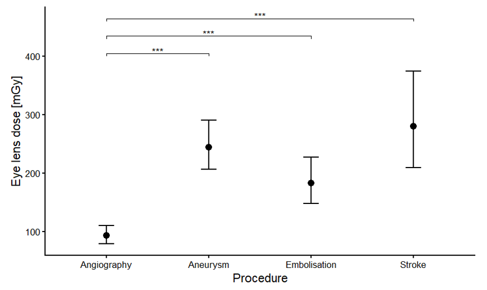
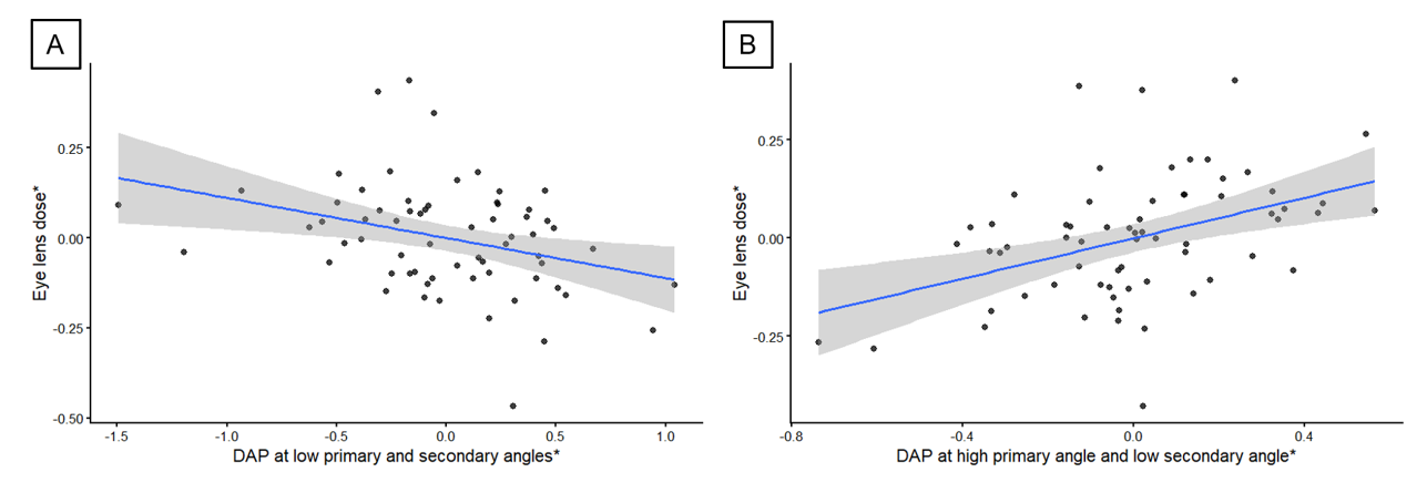
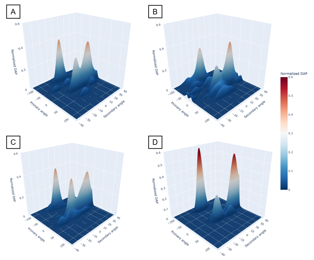
Outlook & Discussion
The regression findings are consistent with the established higher eye-lens exposure from lateral irradiation, while under-table irradiation was associated with lower eye-lens dose when accounting for the other irradiation components. The distinct Stroke angulation profile may partly explain its numerically higher eye-lens dose and indicates systematic procedure-specific differences in irradiation geometry relevant to eye lens exposure. Although projection angles are clinically determined, angulation information could support procedure-specific eye-lens dose estimation, monitoring, and identification of examinations in which closer attention to patient eye-lens exposure may be warranted.
Reference
[1] NCRP Report no. 168
Objective
Diagnostic radiation exposure varies due to automatic exposure control, patient size, scan length, examination complexity and acquisition parameters, which can obscure protocol-, hardware-, or software-related changes. This study determined the protocol-specific sample sizes needed to detect dose reductions in computed tomography (CT), mammography and fluoroscopy/angiography.
Methods
Diagnostic dose data exported from OpenREM were analysed. Protocols with known or suspected changes were excluded to include only stable acquisitions. Metrics included CTDIvol and DLP for CT, average glandular dose (AGD) for mammography, and dose area product (DAP) and dose at the reference point (DRP) for fluoroscopy/angiography. Simulated distributions were generated by reducing values by 0–30%, with 10–100 patients per group to mimic dose optimization. For each group, 4000 Monte Carlo iterations were performed. Log-transformed samples were compared using Welch t-tests and bootstrap and permutation analyses of means and medians, with 2000 resamples. Power was defined as the proportion detecting a change at p < 0.05; 95% power was used as the target.
Results
Required sample sizes depended on protocol variability, dose metrics and reduction magnitude.CTDIvol was less variable than DLP. For non-contrast head CT, a 20% reduction required 30 patients for 95% power. For thoracic CT, a 20% reduction required 100 patients. For mammography AGD, a 20% reduction required 30 patients. For neurointerventional and coronary angiography with percutaneous coronary intervention, a 30% reduction required 55–60 patients using total DAP and DRP. Real clinical CT data before-and-after optimization of a urinary stone CT protocol were analysed: following protocol optimisation, CTDIvol decreased by 27.4% and DLP by 26.2%, and logscale bootstrap mean analysis achieved 95% power at 40–45 patients per group. Mean-based analyses were more robust for highly variable protocols than median-based analyses. Bootstrap analysis on the log scale performed consistently across modalities and agreed with Welch and permutation approaches.
Outlook & Discussion
Protocol-specific sample sizes are necessary to distinguish meaningful dose changes from variability. Log-scale bootstrap analysis provides a robust evaluation method. Dose management systems supply the large datasets required for quantitative protocol optimisation and long-term clinical dose monitoring.
Purpose
Reliable dose management depends on complete examination data, accurate device identification, and alerts that reach the appropriate clinical teams. Guided by IAEA and ESR EuroSafe Imaging recommendations, we extended the open-source system OpenREM with nuclear medicine (NM) data integration, retrospective data quality control, personalized alerts, effective-dose estimates, and additional CT and fluoroscopy metrics.
Methods and materials
OpenREM’s software dependencies and deployment were updated. To address incomplete PACS query responses, OpenREM can recover metadata from a minimally retrieved dose report or other suitable DICOM objects. New overviews compare queried studies, accession numbers, Patient IDs, and stations with records already imported into OpenREM. Devices can be classified by monitoring priority across query and workload views. Alert routing supports recipients for global thresholds, optionally restricted to a device, and personalized rules combining device, intervention, threshold, and recipient.
Results
The retrospective workflow helps medical physicists identify missing examinations, incomplete metadata, and devices requiring review. Query details show whether DICOM objects were retrieved for dose import, metadata recovery, or CT data enrichment; staged DICOM headers can be inspected directly. Filters, record counts before loading, and consolidated exports support review of large datasets. Device examination counts and workload can be analysed using the shared classification. NM examinations can now be included in dose management, while effective-dose estimates provide an additional metric. CT irradiation events can be linked to reconstructed images through the Irradiation Event UID, adding available series descriptions, image-quality information, and reconstruction parameters to exports. Fluoroscopy views include skin-dose filters and skin dose computation. Personalized alert rules allow teaching or procedure-specific thresholds to notify the responsible recipient while retaining general clinical thresholds.
Conclusions
OpenREM now offers practical tools to check data completeness, manage devices, and direct dose alerts according to clinical responsibilities. As an open-source alternative to commercial dose management systems, it allows local workflows and analyses to be adapted and its data processing to be inspected. These developments support radiation protection and quality assurance across imaging modalities.
Background
Swiss fluoroscopy shielding calculations use weekly current–time product (mA·min) as a workload parameter. However, beam filtration alters air kerma per mAs, potentially weakening the link between electrical workload, patient dose indicators, and patient-scattered radiation relevant to structural shielding. We compared matched Siemens Artis and Philips Allura angiography systems.
Methods
Data from four matched device pairs were retrieved via OpenREM, which reads the corresponding RDSRs: neuroangiography, body angiography, and two cardiac laboratories. For each patient procedure, we analysed current–time product, dose–area product (DAP), reference-point air kerma (DRP), copper filter thickness, and tube voltage. For each device, mean DAP and kerma were divided by mean mAs; Siemens/Philips ratios of these quotients were calculated. Radiation output per mAs was additionally modelled with SpekPy for 80-kV tungsten beams with 2.5 mm Al baseline filtration and device-mean filters: Siemens, 0.71 mm Cu; Philips, 0.34 mm Cu plus 1.0 mm Al.
Results
Siemens systems had higher current–time products per procedure in every pair. Siemens/Philips current–time-product ratios were approximately 1.8 for neuroangiography, 4.6 for body angiography, and 2.0 and 1.7 for the cardiac laboratories.
Nevertheless, Siemens systems showed lower DAP and reference-point air kerma per mAs. Siemens/Philips DAP-per-mAs ratios were 0.59, 0.48, 0.80, and 0.87; corresponding reference-point-kerma-per-mAs ratios were 0.56, 0.36, 0.60, and 0.59. Siemens operated at lower mean tube voltages (72/78, 71/75, 81/87, and 83/87 kV, Siemens/Philips) and used thicker copper filters (device means 0.67–0.78 versus 0.29–0.38 mm). SpekPy calculated a Siemens/Philips air-kerma-per-mAs ratio of 0.47, within the clinical reference-point-kerma-per-mAs range (0.36–0.60).
Conclusions
Higher Siemens current–time products do not translate into proportionately higher recorded DAP or reference-point kerma. Filtration plausibly explains part of this discrepancy, although voltage, field size, and clinical technique may also contribute. Current–time product alone may therefore overstate differences in shielding workload between angiography systems. A weekly DAP-based calculation accounting for beam quality, scatter geometry, and barrier transmission should be compared with the Swiss method. In existing rooms, long-term passive area dosimetry at selected occupied locations could assess exposure under documented workloads and independently check shielding adequacy.
Objective Proton therapy assumes a constant relative biological effectiveness (RBE) of 1.1, although RBE varies with factors including linear energy transfer (LET), which increases towards the end of the proton range. This motivates consideration of radiation quality in treatment planning to limit high-LET contributions in healthy tissue. The recently proposed dirty-dose concept, defined as the dose deposited by individual protons at LET above a specified threshold, translates LET information into a dose-based quantity that can be directly incorporated into treatment planning.
Establishing whether calculated dirty dose corresponds to experimentally distinguishable differences in radiation quality could provide a physical basis for its relevance as a treatment planning quantity. This study investigates whether dirty dose can be experimentally assessed using passive optically stimulated luminescence (OSL) detectors.
Methods OSL measurements at two institutions used a single-field spread-out Bragg peak sampled at different depths (I1) and two-field plans with different homogeneous dirty-dose fractions (I2), together covering the relevant dirty-dose fractions. Dirty-dose fractions, defined as the fraction of total dose from individual protons with LET > 3.0 keV/µm, were calculated using the Monte Carlo code SHIELD-HIT (I1) and the treatment planning system RayStation (I2). During readout, the irradiated films were optically stimulated to measure signals from two emission bands with different responses to radiation quality. Their ratio was evaluated against the calculated dirty-dose fraction.
Results Dirty-dose fractions ranged from (4.3–93.2) %, with corresponding OSL signal ratios 1.9–3.1. The dirty-dose fraction followed a logistic dependence on the luminescence signal ratio within experimental uncertainties. This relationship enables quantitative estimation of the dirty-dose fraction from the detector response without prior knowledge of the irradiation field, converging towards dirty-dose fractions 0 and 1 at low and high OSL signal ratios, respectively.
Outlook & Discussion The results support the experimental measurability of dirty-dose fraction and highlight the potential of passive luminescent detectors for its assessment. Further validation could establish these detectors for experimentally evaluating dirty-dose-based planning strategies, supporting the clinical transition towards radiation-quality-aware treatment planning in proton therapy.
Objective. Ultrashort-pulsed electron beams (UPEBs) are increasingly studied in radiobiology and for potential radiotherapy applications, but the time course of normal-tissue responses remains incompletely characterized. Because oxidative injury contributes to radiation-induced central nervous system damage, defining recovery kinetics may inform normal-tissue risk assessment. We characterized post-irradiation redox and enzymatic responses in rat brain after low-energy laser-driven UPEB whole-body exposure.
Methods. In two independent experiments, 56 male Wistar rats were assigned to control, sham-anesthetized, and irradiated groups (n=8 per post-irradiation time point). Whole-body irradiation was delivered at AREAL (3.6 MeV electrons, 450 fs pulses, 2 Hz) to 2 Gy. Brain tissue was examined on days 1, 3, 7, 14, and 28. Oxidative stress was assessed by malondialdehyde (MDA) and protein carbonyl derivatives; superoxide dismutase (SOD), catalase (CAT), creatine kinase (CK), purine nucleoside phosphorylase (PNP), and creatine were measured as antioxidant, metabolic, and immune-related markers.
Results. MDA more than doubled by day 1 and peaked on day 3, while protein carbonyl derivatives increased threefold or more on day 3; both returned to control by day 28. SOD activity decreased during days 1–7, partially recovered by day 14, and returned to control by day 28. CAT remained near control early and showed an almost twofold compensatory increase on day 14 before normalizing by day 28. CK activity fell by more than 20% on day 1, remained reduced through day 7, and returned to control on days 14–28; creatine transiently increased on day 3. PNP showed a time-dependent response, although interpretation was limited by anesthesia-related inhibition.
Outlook & Discussion. Acute UPEB exposure produced a coordinated, largely reversible redox and metabolic response in brain tissue. The response kinetics paralleled the reported acute phase of radiation-induced brain injury, with substantial recovery during weeks 2–4. These time-resolved normal-tissue data are relevant to emerging pulsed electron-beam modalities. Direct comparison with conventional electron irradiation is needed to determine which effects are specifically attributable to ultrashort pulse structure.
Objective: Current cancer treatment modalities often fail to achieve complete tumor eradication. Over the past two decades, our group at the Institute of Anatomy at the University of Bern, has investigated the radiobiological mechanisms underlying sub-millimeter Spatially Fractionated Radiotherapy (smSFRT), an innovative radiotherapy approach that delivers high tumoricidal radiation doses while sparing normal tissues, with the goal of enabling its clinical translation and improving outcomes for cancer patient.
Methods: We investigated the biological effects of smSFRT in multiple preclinical models, including murine melanoma (microbeams of 50 μm width, spaced 400 μm um centre-to-centre) and the chick embryo chorioallantoic membrane (CAM) (minibeams of 500 μm width, spaced 1100 μm centre-to-centre). More recently, we initiated the first veterinary clinical trial of smSFRT at the Australian Synchrotron in Melbourne (microbeams of 50 μm width spaced 400 μm centre-to-centre). Across these models, vascular responses and immune effects were characterized at multiple time points following irradiation. In parallel, since 2024 we have contributed to the first human clinical trials of smSFRT, specifically minibeams, in the United States.
Results: We demonstrated that smSFRT controls melanoma by eliciting an antitumor immune response that enhances antigen presentation and cytotoxic T-cell activation, and that it synergizes with immunotherapy. Using clinically relevant irradiation parameters (dose and dose rates), we showed that smSFRT induces transient, reversible vascular permeability in the CAM, supporting improved drug delivery. Furthermore, smSFRT achieved promising therapeutic responses both in spontaneous human-sized canine tumors and in the first clinically treated patients, supporting its translational potential.
Conclusions: Our findings establish a strong biological basis for the clinical translation of smSFRT. By promoting robust antitumor immunity, synergizing with immunotherapy, and transiently increasing vascular permeability to facilitate drug delivery, and combined with its well-established normal tissue sparing, smSFRT represents a promising multimodal strategy for improving the treatment of resistant cancers.
Objective
Pancreatic ductal adenocarcinoma (PDAC) is characterized by a hypoxic microenvironment that
contributes to resistance to radiotherapy. While standard clinical monitoring relies on
macroscopic changes in tumor size, such lagging indicators fail to capture early physiological
shifts in oxygenation and vascular remodeling. We implemented a multiparametric imaging
approach to characterize these responses and identify interpretable early response markers.
Methods
A co-registered 3D imaging platform integrating volumetric multispectral optoacoustic
tomography (MSOT) and functional magnetic resonance imaging (MRI) was used to characterize
acute radiotherapy responses in human PDAC xenografts. Twenty mice bore subcutaneous
hindlimb tumors (10 MIA PaCa-2, 10 SU.86.86). Ten mice received a single 10 Gy dose, and 10
served as sham-irradiated controls (0 Gy). Tumors were monitored over five consecutive days,
beginning before irradiation. Voxel-wise fitting of oxygen-challenge responses with a modified
logistic function yielded volumetric maps of blood oxygen saturation, oxygen-induced changes
in saturation, and response kinetics. Fits to variable flip angle MRI scans yielded
complementary tissue oxygenation-sensitive longitudinal relaxation rate (R1) measurements.
Delta radiomic features extracted from depth-stratified profiles were analyzed using partial
least-squares regression and variable importance scores.
Results
Irradiated tumors exhibited increases in blood oxygen saturation consistent with vascular
reoxygenation. Depth-resolved analysis captured spatial heterogeneity in these responses.
Partial least-squares analysis identified patterns distinguishing irradiated tumors from controls,
with features reflecting peripheral oxygenation, oxygen-enhancement magnitude, and
heterogeneity contributing prominently. MRI provided complementary anatomical and oxygensensitive information for multiparametric tumor characterization. MSOT-derived features
revealed more pronounced treatment-associated patterns than R1-derived features, suggesting
that MSOT may be more informative for detecting early functional changes.
Outlook & Discussion
This platform provides a framework for identifying interpretable early radiotherapy response
markers and characterizing potential windows of tumor reoxygenation. The findings support
further development of oxygenation-guided adaptive fractionation and personalized
radiotherapy in pancreatic cancer. Validation against long-term treatment outcomes is needed
to establish the prognostic value of these markers.
Objective
Ultra-high-dose-rate (UHDR) radiotherapy requires prospective clinical evidence for assessment of its feasibility, safety and clinical benefit of the Flash-effect. Accurate dose monitoring and reporting remain major challenges. This work presents the clinical application of in-vivo dosimetry and RTQA program in the phase I Flash-Skin I trial (NCT06549439), the first clinical study of fractionated electron Flash-RT delivered on a modified conventional C-arm linac.
Methods
Seven patients with metastatic melanoma and cutaneous or sub-cutaneous metastases were treated with 2x9 Gy UHDR electron fractions followed by 1x9 Gy conventional electron radiotherapy using a modified Varian TrueBeam (9 MeV, 1.08 Gy/pulse, 216 Gy/s average). A dedicated RTQA program was implemented and validated over 15 months before patient treatments. In-vivo dosimetry combined radiochromic films, OSLD, plastic scintillators and diamond detectors. OSLDs were calibrated against a reference ionization chamber and used for dose reporting, while active detectors provided pulse-to-pulse and intra-pulse measurements.
Results
OSLD measurements agreed with the reference chamber within ±2.9% (k=1), establishing OSLDs as the primary dose-reporting system. For the 14 treated UHDR fractions pulse-to-pulse and intra-pulse stability were within 3.8% (min-to-max) and 2.8% (std), respectively. Film dosimetry confirmed agreement between planned and delivered dose distributions (gamma pass >95%, 5%/3mm). All seven patient treatments were successfully delivered with maximum dose deviations of +4.6% per fraction and +2.8% for total delivered dose. Clinically, no dose-limiting toxicity occurred. Five patients developed grade I-II radiation dermatitis and one reported grade I pain. Local responses included one complete remission, three partial responses, and three stable lesions.
Outlook & Discussion
The proposed multi-detector approach enabled accurate dose verification, temporal beam monitoring, and dosimetric redundancy during clinical UHDR delivery. Together with the successful implementation of the RTQA program and the clinical outcome of the Flash-Skin I trial, these results demonstrate clinical feasibility and safety of Flash-RT at a converted C-arm linac, providing a framework for further future UHDR clinical trials and broader clinical implementation
Objective: Modelling and predicting cellular radiosensitivity has been of interest in radiation biology for decades. In this preliminary and exploratory work, an analysis of radiation dose-response and transcriptomic data is performed for 14 canine oral malignant melanoma cell lines. The objective of this analysis is to explore the correlations between gene expression and intrinsic radiosensitivity.
Methods: In a first step, a pre-existing radiosensitivity model was applied to the canine data. In a second step, a gene- and pathway-level correlation analysis with four radiobiological parameters $\text{SF2}$, $\text{AUC}_\text{log(SF)}$, and fitted $\alpha$ and $\beta$ from the Linear-Quadratic model was performed.
Results: The published model could not be applied satisfactorily to the canine dataset, as only 3 of the 14 cell lines showed an agreement within 10% between prediction and measurement. The genome-wide analysis led to no significant correlation between gene expression and radiobiological parameters. Five DNA repair pathways were also selected for correlation analysis, and non-homologous end joining was found to be significantly correlated to both alpha and beta.
Outlook & Discussion: The unsuitability of the radiosensitivity model to the canine cell lines might originate in part from the difference in transcriptome profiling methods. Moreover, the limitations of this model have been investigated more closely in the recent years. The difference between the number of cell lines and dimension of the transcriptome vector in this analysis is of several orders of magnitude (14 vs $2\times 10^4$). It is then not surprising that a simple correlation analysis could not yield any significant result. Pathway analysis allows for aggregation of gene expression across predefined gene sets, thereby reducing the size of the feature space. The observed correlation of the NHEJ pathway with both alpha and beta appears promising and is biologically plausible given the established role of NHEJ in the DNA repair. To strengthen this finding, the association should be further investigated in other datasets. Future work should combine increased sample sizes with biologically informed, targeted analyses to reduce the search space.
Objective
In 2004, the president of SSRMP asked whether our society, founded in 1964 as the Swiss Society for Radiation Biology, had lost its basis. At SASRO 2026 I showed that Swiss radiation biology was in fact restructuring after its 40-year-old institute in Zürich, home to the only Swiss chair of radiation biology, became a cancer-research institute in 2003. For SSRMP we asked two questions: is the field growing or shrinking, and if growing, who trains its researchers?
Methods
With the help of Claude (Anthropic), we compiled a census from public records containing 1) radiation-biology journal papers with a Swiss author, 1950–2026 (708 full papers); 2) Swiss doctoral theses in radiation biology, radiation oncology, medical physics and related fields, with their supervisors; and 3) where Swiss-affiliated authors with a public ORCID profile earned their doctorate.
Results
Radiation-biology papers rose from about one a year in the 1960s to about 40 a year since 2020. Most of the growth since the 2010s comes from FLASH and Spatially Fractionated Radiotherapy (almost half the papers since 2020). Training grew far more slowly: from the 1960s to the 2020s, radiation-biology doctorates only doubled (15 to 31 per decade). Medical-physics doctorates, by contrast, rose almost six-fold in a single decade (from 10 in the 2000s to 57 in the 2010s). The largest supervisor in the records is Martin Pruschy in Zürich (23 radiation-biology doctorates since 2002). Much of the recent research workforce trained abroad: six in ten authors active since 2016 whose doctorate could be traced earned it outside Switzerland.
Outlook & Discussion
Have we lost our radiation-biology basis? We argue: no, but we import much of it. One reason we will discuss: radiobiology is required and examined in the training of radiation oncologists, medical physicists and radiation therapists, yet there is no programme, professional title or examination for radiation biologists. We will present the evidence and its limits, and invite discussion on a national radiobiology course.
Objective: Elective Nodal Irradiation (ENI) for head and neck cancer aims to decrease regional recurrence but contributes to side effects. Probabilistic treatment planning based on tumour control probability (TCP) and normal tissue complication probability (NTCP) may improve trade-offs. To that end, a previously introduced model for regional TCP in electively irradiated lymph node levels (LNL) [1] was further developed.
Methods: Each voxel i is assigned a probability qi of harbouring an occult metastasis (OM). TCP is given by TCP=∏i[qiTCPi(di)+(1-qi)], where qiTCPi(di) is the probability of voxel i harboring an OM and being successfully controlled with dose di, and (1-qi) is the probability of voxel i not harboring an OM. qi is defined as a continuous, monotonically decreasing function of distance from the primary tumour, whose parameters are chosen such that the model yields the estimated overall probability of occult metastases in each relevant LNL [2]. To account for motion/setup uncertainty, qi is convolved with a Gaussian. For two oropharyngeal cancer patients, treatment plans were optimized using TCP and NTCP models for xerostomia, dysphagia, or hypothyroidism as objectives.
Results: Compared to standard treatments delivering a homogenous elective dose to LNLs, the probabilistic planning approach may reduce NTCP by compromising coverage of the LNL near organs-at-risk. By incorporating spatially varying qi maps, coverage is compromised preferentially in regions of lower qi, and less when closer to the primary tumour - resulting in only minor reductions in regional TCP.
Outlook & Discussion: The previously introduced model [1] assumed a homogeneous qi distribution within a PTV around each LNL. This resulted in unrealistic abrupt changes at the boundaries between LNLs, and an inconsistent handling of motion/setup uncertainty. The improved model presented here, contributes to efforts to develop probabilistic methods to incorporate uncertainty in subclinical tumour extension in treatment planning.
[1] Moos, PMB,71(3),2026.
[2] Ludwig, Sci. Rep. 15:18152, 2025.
Objective
Lattice radiotherapy (LRT) delivers heterogeneous dose distributions by placing high-dose vertices within the tumor and is a promising strategy for large tumors. This work aimed to develop a clinically feasible and robust LRT planning strategy for an upcoming clinical trial at USZ.
Methods
Twenty-five patients with large tumors (PTV > 300 cc, long-axis diameter > 10-cm) treated within the last 5 years were retrospectively selected. Plans were generated using 7-mm-radius spherical vertices within the GTV cropped 10-mm from OARs (Boost GTV), a PTV periphery dose of 20 Gy in 5 fractions, and vertex doses above 50 Gy. Center-to-center spacings of 30 and 40-mm were compared for vertex dose, valley dose, peak-to-valley dose ratio (PVDR), dose spills, and OAR sparing. Robustness against systematic and random interfractional setup errors was assessed by simulating isocenter shifts over five fractions. Additionally, a 7-mm margin between vertices and GTV boundary, in union to the 10-mm OARs margin, was investigated to provide space for dose fall-off.
Results
Clinically acceptable LRT plans were achieved for all patients. Compared with 30-mm spacing, 40-mm spacing achieved higher PVDR, lower valley dose, reduced dose spill, and similar or lower OAR doses, with these advantages maintained under setup uncertainties. However, larger spacing reduced vertex number. As dosimetric benefits were more pronounced in large tumors, 40-mm spacing was selected for Boost GTV > 200 cc and 30-mm spacing for smaller tumors, balancing spatial dose modulation and vertex number. Nine patients fell into the small-tumor category and 16 into the large. The additional 7-mm GTV margin reduced dose spill under setup uncertainties by maintaining the high-dose gradient within the tumor.
Outlook and Discussion
A clinically feasible and robust LRT planning workflow was developed. Vertex doses were comparable to previous studies, while dose spill and OAR sparing were further evaluated. Based on these findings, a tumor size-dependent lattice geometry was introduced, aiming to preserve spatial dose modulation throughout fractionated treatment while maintaining sufficient vertex number and substantial dose escalation.
Objective
Hypoxia drives radioresistance in solid tumors, while estrogen signaling influences proliferation and treatment response in hormone-sensitive breast cancers. This study developed a 3D agent-based computational model to investigate the combined effects of estrogen exposure, microenvironmental hypoxia, and fractionated radiotherapy in breast cancer spheroids.
Methods
A 3D spheroid model was implemented in PhysiCell. Oxygen diffusion and cellular consumption were modeled to generate spatio-temporal hypoxia gradients toward the core. Estrogen signaling was incorporated as an increased probability of cellular proliferation. Radiation response was simulated using the Linear-Quadratic model (\alpha = 0.30\text{ Gy}^{-1}, \beta = 0.03\text{ Gy}^{-2}), incorporating hypoxia-induced radioresistance via an Oxygen Enhancement Ratio (OER) tied to local oxygen tension. Four conditions were evaluated: control, estrogen exposure, standalone irradiation, and combined estrogen plus irradiation.
Results
The framework captured single-cell spatial dynamics, localized oxygen depletion, and treatment-induced cell death. Estrogen exposure accelerated overall spheroid growth and expanded the central hypoxic core. Hypoxia-mediated radioresistance blunted cell killing within inner spheroid regions during fractionated delivery. In the combined condition, estrogen-driven proliferation accelerated inter-fraction repopulation, partially counteracting radiation-induced cell death and altering net treatment response compared to non-estrogen controls.
Conclusion
This model provides a mechanistically grounded platform to study interactions between hormonal signaling, tumor microenvironmental hypoxia, and radiotherapy response in breast cancer spheroids. While experimental validation against 3D in vitro data is necessary to calibrate parameters, the framework highlights how hormone-driven proliferation and microenvironmental gradients jointly influence fractionated radiotherapy outcomes.
Objective: Online adaptive radiotherapy (oART) requires adjusting treatment plans to daily anatomy within minutes. Monte Carlo (MC) beamlet calculation is time-consuming. We hypothesize that target and organ-at-risk (OAR) similarity between daily and earlier anatomies can guide selection of pre-calculated MC beamlet atlases. We test this and quantify resulting plan quality and computation time.
Methods: MC beamlets pre-calculated on earlier fractions (atlases) were reused to optimize VMAT plans on the daily anatomy, followed by final MC dose calculation. Similarity between daily and atlas anatomies was quantified for PTV and OARs (DSC, MSD, HD95). One lung (Dp=60 Gy; two fractions, three of four atlases each) and one esophagus case (Dp=50.4 Gy; three fractions, two of six atlases each) were adapted. Dose-volume differences between plans with reused and daily recalculated beamlets were related to PTV and OAR similarity, and beamlet preparation times (recalculation vs. atlas selection and reuse) were compared. HI was defined as (D2%−D98%)/Dp.
Results: In one lung fraction, the atlas with the highest PTV similarity performed worst of three atlases, while the atlas with the closest heart similarity performed best. Worst vs. best: PTV DSC 0.78 vs. 0.63, heart MSD 3.7 vs. 2.5 mm, PTV D99% −3.1 vs. −1.7 Gy, HI 0.108 vs. 0.079 (recalculated 0.056), heart D5% +3.9 vs. +2.2 Gy. In esophagus, three fractions with similar PTV agreement to the same atlas (MSD 3.5–3.8 mm) ranged from PTV D99% −0.5 to −11.2 Gy, with OAR D5% differences of 2.9–7.1 Gy. With the best-matching atlas, fractions 15 and 27 stayed within 1.2 Gy for all dose metrics. Beamlet preparation time decreased by 52% (esophagus) and 18% (lung).
Outlook & Discussion: PTV similarity alone was insufficient: in one of four quantified multi-atlas comparisons it selected the worst, whereas the best atlas always had the closest heart similarity. OAR similarity should therefore complement target similarity in atlas selection, pending validation on larger cohorts and a speed-optimized implementation.
Supported by SNSF grant 10001696 and Siemens Healthineers through their business line Varian.
Objective
Stereotactic radiosurgery is established for trigeminal neuralgia (TN), where dose optimization and accuracy are critical near small, high-risk targets. This study compares plan quality and delivery efficiency between RayStation and Precision for CyberKnife TN radiosurgery.
Methods
Ten patients previously treated on a CyberKnife S7 with Precision 3.5.0.1 were retrospectively re-planned on RayStation 2024A SP1 using a fixed 5 mm collimator and identical contours/prescription. Plans underwent patient-specific QA (SRS MapCheck, SunNuclear; 5%/1 mm gamma criteria, 10% threshold). Metrics compared: gradient index (GI), conformity index (nCI), monitor units (MU), segments, nodes, delivery time, and maximum dose (D0.03cm3) to brainstem, cranial nerve (NCV), and gasserian ganglion (GGl; D0.01cm3), via Wilcoxon signed-rank exact test.
Results
All plans passed PSQA (mean gamma pass rate at 5%/1 mm: RayStation 99.6%, range 97.8–100%; Precision 99.1%, range 95.8–100%). RayStation showed a lower mean GI (8.1 vs. 8.8, p=0.03) and higher mean nCI (2.31 vs. 2.21, p=0.69). Maximum NCV dose was significantly lower with RayStation (mean 3.1 ± 1.3 vs. 4.6 ± 1.5 Gy; median 3.1 vs. 4.9 Gy; p = 0.04) but values remain well below any toxicity threshold and therefore have no expected clinical consequences. BS Dmax did not differ significantly (10.6 ± 1.5 vs. 10.7 ± 1.8 Gy; median 11.1 vs. 10.5 Gy; p = 0.92), as was GGl Dmax in 9 evaluable pairs (mean 22.3 ± 12.5 vs. 24.9 ± 11.1 Gy; median 26.1 vs. 20.4 Gy; p = 0.36) and total MU (19194 vs. 18675). Precision required more segments (85.8 vs. 73.2) and nodes (51.5 vs. 42.9) but achieved a shorter delivery time (2652 vs. 2861 s).
Outlook & Discussion
Both TPS produced clinically acceptable, PSQA-validated plans; NCV Dmax was the only significant difference, and given the small sample, other trends remain exploratory.
Conclusion
Both TPS generated PSQA-validated plans meeting institutional constraints. RayStation showed a trend toward steeper gradient and lower NCV dose, at the cost of a slightly longer estimated delivery time. Confirmation in a larger cohort is needed.
Objective
In small-field radiosurgery (5 mm cone, 100 Gy), active detectors lack tissue equivalence at the measurement point, requiring complex cavity theory, simulations and output corrections — despite offering seamless electrical readout. Conversely, Gafchromic EBT-XD film generates a signal directly proportional to dose in a tissue-equivalent environment, providing a true 2D "WYSIWYG" image. However, film dosimetry shifts complexity to the downstream evaluation chain (scanner optics, optical density resolution, calibration). This study establishes an extended-range (up to 100 Gy) 2D film protocol by controlling the optical readout for stereotactic radiosurgery (SRS) plan verification.
Methods
Trigeminal SRS plans (5 mm cone, 6X-FFF, Varian TrueBeam) were delivered in a CBCT registered phantom. To control the evaluation chain, films were digitized using a high-OD scanner (Epson V750 Pro, 4.0 DMax) with triple-channel (RGB) analysis (FilmQA Pro). Absolute dose normalization relied on a direct 5-level calibration curve (0, 10, 20, 40, 100 Gy). Film dose maps were evaluated without software spatial shifts. Parallel point measurements used a PTW microDiamond detector, corrected via IAEA TRS-483 (factor 0.962).
Results
High-OD scanning and dynamic RGB weighting prevented optical saturation at 100 Gy. Unshifted film QA achieved excellent spatial-dosimetric agreement, with Gamma passing rates of 99.9% (Case 1) and 99.8% (Case 2) at 3%/1mm. Conversely, the microDiamond detector showed systematic negative deviations (-6.7% and -4.8% post-TRS-483 correction), reflecting active volume averaging (~2 mm) and angular dependence in 5 mm fields within the -7% clinical tolerance.
Outlook & Discussion
While active probes offer simple signal transmission at the expense of cavity perturbations in minute fields, radiochromic film provides true tissue-equivalent 2D verification when the optical evaluation chain is rigorously managed. This 5-level, high-OD protocol eliminates lengthy linac recalibration time, delivering a spacially and dosimetrically accurate QA solution for up to 100 Gy dose SRS
Objective:
Monte Carlo simulations for submillimeter spatially fractionated radiotherapy (sub-mm SFRT) are computationally expensive due to high spatial resolution, accuracy and precision requirements. To address this issue, a linear regression model (LRM) was developed predicting optimal voxel sizes (OVSs) aiming to reduce the required computational resources whilst minimizing compromise in dosimetric endpoints.
Methods:
Ground-truth dose distributions in water (1×1000×1000 μm3 voxel size) were calculated using the Swiss Monte Carlo Plan (TrueBeam 6 MV photon beam, 8 cm tungsten multislit-collimator) for beamwidths (BW) in the range of 0.5-1.5 mm and center-to-center spacings (CTC) of 1.0-18.0 mm. Ground-truth crossplane profiles were fitted using superimposed Pseudo-Voigt functions (PVF), which subsequently served as reference profiles. The PVFs were integrated over equally large sub-intervals ranging from 25-1000 µm to represent the dose in the voxels of corresponding size in crossplane-axis. Peak-to-valley dose-ratio, mean peak and mean valley dose degradation resulting from increasing voxel sizes was quantified w.r.t. the ones on the reference profile. OVS for each geometry was defined as the largest sub-interval resulting in <1% metric deviation from the reference. The LRM was then created to predict OVS across the investigated BW and CTC-ranges. The LRM was evaluated on a clinically motivated brain cancer case (BW of 0.52 mm and CTC of 6.26 mm) by comparing the previously mentioned dose metrics from a highly resolved simulation with the ones obtained using the LRM-predicted OVS at three different depths.
Results:
The OVSs ranged between 131.1-222.9 µm for the investigated geometries. The LRM resulted in OVS = 91.75 µm/mm x BW + 82.25 µm. The dosimetric metrics agreed within 1.3% in the clinically motivated case. With the predicted OVS, the computational time and memory can be reduced by a factor of approximately 4-5 relative to previously used settings.
Outlook & Discussion:
We successfully developed a LRM predicting OVS for sub-mm SFRT. This model has the potential to reduce computational costs for Monte Carlo dose calculations in sub-mm SFRT.
Supported by SNSF grant 10002984
Aim: Thermal dose, expressed as cumulative equivalent minutes at 43°C (CEM43), is considered a predictive parameter of treatment response to combined radiotherapy (RT) and hyperthermia (HT). Thermometry data from locally advanced rectal cancer (LARC) patients enrolled in the Hyperboost REC study were used to assess the spatial and temporal variability of CEM43 thermal dose metrics.
Methods: In total, 206 LARC patients treated with neoadjuvant chemoradiotherapy (CRT) and HT (median age: 62 years, male 66%) were included in this analysis. RT was given with a median dose of 50.4Gy (95%CI:50.4─56) combined with 5-FU only (46% of patients). The median number of HT sessions was 7(1-11). Treatment data files from the BSD-2000 3D/MRI system (Pyrexar Medical Corporation, USA) were used to evaluate thermal dose metrics: CEM43T10, CEM43T50, and CEM43T90. Temporal thermometry was based on a single measurement point (i.e., the tip of the temperature probe inserted in rectum during HT) recorded every 5 seconds, whereas spatial mapping was derived from temperature measurements along the catheter length in 1-cm increments up to a maximum depth of 10 cm, acquired at 5- or 8-minute intervals.
Results: There was no significant difference between spatial and temporal evaluations of the thermal dose parameters CEM43T10 and CEM43T50. However, significant variability was observed for the low-temperature parameter CEM43T90. Subgroup analyses stratified according to the median number of HT sessions revealed similar results for spatial and temporal evaluations. Furthermore, the analysis according to treatment period (before versus after 2015) showed that patients treated after 2015 received higher CEM43T10 thermal doses than those treated before 2015 (5.29±7.88 vs 3.66±3.19, p=0.03), while CEM43T50 and CEM43T90 were comparable between groups. Patients who had complete responses tended to receive higher thermal doses (CEM43T50>1.3 min and CEM43T10>3.4 min) and more HT sessions (>7).
Conclusion: This analysis showed that the type of thermometry influences the evaluation of CEM43T90 metric. A trend toward complete response was observed in patients who received both higher thermal doses and a higher number of HT sessions.
Objective: Hypofractionation can reduce treatment burden and resource use in radiotherapy, but increasing dose per fraction may increase normal tissue complication probability (NTCP). Conversely, improved geometric precision may enhance organ-at-risk sparing and compensate for this increase. Using head-and-neck (H&N) proton therapy as a test case, we investigated whether and to what extent fractionation could be reduced while maintaining the NTCP of a conventionally fractionated reference treatment.
Methods: Four H&N proton therapy patients are analysed retrospectively. A clinically plausible 33-fraction plan with 3-mm setup uncertainty is used as reference. Hypofractionated plans are generated with fraction doses up to 5.5 Gy (RBE), with prescription and optimization doses adjusted to match the reference EQD2. Setup uncertainties of 3, 2, 1, and 0 mm are investigated. Target coverage (mean dose and V95%) and xerostomia NTCP are evaluated. For each setup uncertainty, the minimum number of fractions maintaining NTCP at or below the reference value, at comparable target coverage, is identified. Time saving is estimated, assuming a treatment time of 30 minutes per fraction.
Results: At a given setup uncertainty, NTCP increased as the number of fractions decreased. In contrast, reducing setup uncertainty lowered NTCP. The balance between these opposing effects determined the maximum degree of hypofractionation achievable without exceeding the reference NTCP. With a 1-mm setup uncertainty, treatment could be reduced from 33 to a mean of 20 fractions (range, 10–25), while maintaining comparable target coverage. This corresponds to an average reduction of 13 fractions, or 390 treatment minutes per patient.
Outlook & Discussion: These results illustrate a general trade-off between fractionation and geometric precision: improved uncertainty management may create room for hypofractionation without increasing predicted normal-tissue toxicity. In H&N proton therapy, online adaptive workflows may provide the geometric precision required to approach 1-mm uncertainty, potentially enabling shorter treatment courses and lower overall resource use.
Objective
In conventional ocular proton therapy, surgical fiducials (clips) are sutured onto the sclera around the tumor for eye modeling and patient positioning. Target-beam alignment is verified using in-room X ray imaging and iterative manual matching of the clips to their planned locations. The present work investigates the accuracy and the clinical impact of a noninvasive positioning approach, based on anatomical eye modeling and optical pupil tracking.
Methods
Patient-specific eye models of 17 patients with ocular cancer treated with the conventional approach were derived from MRI as geometrical approximations of the underlying anatomy. During positioning, the patient’s head was fixed, and gaze directed to a fixation light while X-ray images were acquired. Simultaneously, two calibrated optical cameras recorded pupil images. Eye pose was defined by two rotation angles (azimuth, elevation), and 3D position of the eye center. Using the eye models, pupil contours were projected onto the camera images for a range of eye poses. Light refraction through the cornea and the aqueous humor was accounted for. An optimization routine then determined the eye pose. The approach was evaluated against the clip-based gold standard in terms of accuracy (clip-to-clip deviations) and clinical impact (simulated positioning on the planned dose distribution).
Results
The deviation between tracking-based and clip-based measurements of clip positions was centered around zero with a normative median deviation of 1.12 mm (IQR: 1.07 mm). In a subset of seven cases with complete dosimetry analysis, simulated tracking-based positioning had no dosimetric impact (D98 > 99[RV1.1]%) in six cases; one case showing a notable deviation (D98 = 85%).
Outlook & Discussion
The proposed approach exhibited no systematic bias and was on average comparable to the gold standard. However, the presence of outliers indicates that its precision is limited in a subset of cases, to an extent that may adversely affect clinical accuracy. Future integration of fringe projection profilometry could help address this limitation, serving for validation and real-time positioning correction. Further evaluation on a larger patient cohort is pending.
Objective:
To perform a dosimetric comparison of photon volumetric modulated arc therapy (VMAT) versus static proton arc (SPA)—a novel delivery method utilizing discrete, stationary proton gantry angles—for lattice radiation therapy (LRT) using sequential and simultaneous integrated boost (SIB) techniques.
Methods:
Clinical VMAT plans from ten previously treated patients—seven sequential (3 to 24 spheres) and three SIB (6 to 40 spheres) cases—were compared against retrospective SPA replans. Both modalities deliver a conventional 20 Gy in 5 fractions to the bulky tumor with lattice delivery on top. The sequential approach adds a 20 Gy single-fraction boost to multiple 1.5 cm diameter spheres, minimizing valley doses between them. The SIB approach simultaneously delivers 66.70 Gy to multiple 1.5 cm diameter spheres in 5 fractions, with valley doses constrained to the 20 Gy baseline. Plan quality was evaluated using mean Peak-to-Valley Dose Ratios (PVDR), defined as the ratio of mean dose within high-dose spheres to mean dose of the gross tumor volume (GTV) excluding the spheres. Selected plans were delivered using a Varian TrueBeam linear accelerator and a ProBeam proton gantry to assess clinical delivery times.
Results:
SPA consistently achieved higher mean PVDR than VMAT, with increases of 52.9%–222.1% in sequential plans and 26.4%–42.1% in SIB plans. This was driven by lower valley doses in proton plans, reduced by 32.5%–68.4% in sequential cases, while peak doses remained comparable. SPA maintained high-dose target coverage (95.0%) for SIB plans, whereas VMAT coverage dropped to as low as 66.0%. Delivery times were shorter for VMAT (4.3–7.3 min) than SPA (6.7–16.0 min).
Outlook & Discussion:
SPA provides substantial dosimetric advantages over VMAT for lattice therapy by reducing valley doses and maintaining superior high-dose sphere coverage in SIB plans. These improvements fulfill the primary motivation of lattice therapy: maximizing peak-to-valley dose ratios to safely deliver dose within bulky tumors while minimizing dose to surrounding healthy tissue. Despite longer SPA delivery times, both modalities successfully deliver complex spatially fractionated radiation therapy (SFRT) plans within clinically reasonable timeframes.
Objective
Faraday cups (FCs) could enable dose rate-independent reference dosimetry in proton therapy, including ultra-high dose rate delivery, where ionisation chambers suffer from recombination. The charge collection efficiency of an FC with a suppressor electrode depends on the applied voltage, since the electric field governs the suppression of low-energy secondary electrons emitted from the metal surfaces. It is unclear whether Monte Carlo simulations can reproduce this behaviour, and which physics models are required. We investigated this for the PSI-FC, exposed to a $15\,\mathrm{MeV}$ electron beam at METAS.
Methods
The efficiency was measured from $-1\,\mathrm{kV}$ to $+1\, \mathrm{kV}$. We extended TOPAS with the Geant4-MicroElec models, which provide discrete electron transport down to a few $\mathrm{eV}$ and describe surface emission. Electric field maps were imported, and MicroElec activated in $10\,\mathrm{nm}$ layers at all vacuum-facing surfaces. Four configurations were tested: two using standard physics, with production cuts of $990$ and $50\,\mathrm{eV}$; and two with MicroElec active below $1\,\mathrm{keV}$, using either Penelope or Møller--Bhabha above the threshold. Agreement was quantified with the normalised error $|E_\text{n}|$ at $k=2$, combining Monte Carlo statistics, residual magnetic field and measurement uncertainties; $|E_\text{n}| \leq 1$ indicates agreement within the stated uncertainties.
Results
The measured efficiency was flat at $(98.71\pm0.12)\,\%\; (k=1)$ below $0\,\mathrm{V}$ and fell to $(96.63\pm0.12)\,\%$ at $+1\,\mathrm{kV}$, a step of $2.09$ percentage points ($\mathrm{pp}$). Standard physics produced a step of at most $0.05\,\mathrm{pp}$, regardless of the production cut ($\max|E_\text{n}|= 8.0$). MicroElec combined with Penelope reproduced about half of the measured step ($1.1\,\mathrm{pp}$, $\max |E_\text{n}| = 3.7$), whereas MicroElec with Møller--Bhabha reproduced $95\,\%$ of the measured step ($2.0\,\mathrm{pp}$), agreeing with all measured points ($|E_\text{n}| \leq 0.8$).
Outlook & Discussion
The voltage response of the FC cannot be reproduced by lowering production cuts in condensed-history simulations. Discrete $\mathrm{eV}$-scale transport and surface-emission models are required. The ionisation model above the MicroElec switch-over threshold is also critical, because it sets the electron source that MicroElec transports. Our final model reproduces the FC response within uncertainties, giving a validated physics setup for simulation-based FC studies in reference dosimetry.
Objective
Uncertainties in reference dosimetry for proton radiotherapy are large (1.7%, k=1) compared to conventional radiotherapy modalities. Furthermore, emerging therapy modes delivering ultra-high dose rates challenge the current ionisation chamber (IC)-based codes of practice. We aim to tackle these problems by developing a new pillar of proton reference dosimetry based on Faraday cups (FCs), which are inherently dose-rate independent. By characterising our FC design in detail, and ensuring traceability to primary standards, we intend to establish our FC as a reference instrument for FC-based proton dosimetry.
Methods
At METAS, the national metrology institute (NMI) of Switzerland, we have characterised the entire read-out chain of our designated reference FC. We determined traceable calibration factors for charge measurements using an electrometer, and we quantified the influences of beam characteristics, measurement parameters and environmental conditions on the measurement uncertainty. We also established procedures for measuring the FC’s collection efficiency $\eta$, defined as the ratio between the charge measured by the FC and the total charge delivered by the beamline, in an electron beam at METAS.
Results
Based on our comprehensive characterisation, we estimate an uncertainty of 0.13% (k=1) on absolute charge measurements using our electrometer. We determined the FC's collection efficiency for 15 MeV electrons to be (98.72 $\pm$ 0.12)%, in excellent agreement with Monte Carlo (MC) simulations. The uncertainty contributions from the electrometer and FC efficiency add up to a total uncertainty on electron beam charge measurements of 0.18%.
Outlook & Discussion
In the near future, we will use the procedures developed at METAS to measure the FC’s collection efficiency for clinical proton beams at the Paul Scherrer Institute (PSI), aiming for a similar measurement uncertainty as with electrons. Using MC simulations to convert proton fluence to absorbed dose will then allow us to determine experimental beam quality correction factors $k_Q$ for ionisation chambers and proton beam qualities $Q$ with a target uncertainty of <1% (k=1).
Objective: Macro Monte Carlo (MMC) methods improve dose calculation efficiency over general-purpose MC algorithms by modeling particle transport in large-scale macroscopic steps. So far, MMC has been validated for electron energies up to 25 MeV. This study aims to develop and validate an MMC method for very high energy electrons (VHEE) up to 250 MeV.
Methods: EGSnrc-based simulation modeled electrons incident perpendicularly on spheres (0.2-25 MeV) of 0.5-3 mm radii and slabs (25-250 MeV) of 2 mm thickness of water and human tissue materials. Physical quantities including energy loss, lateral displacement, and angular distributions of primary and secondary particles were scored and stored in a database. This database was subsequently used to sample electron transport in a step-by-step manner, employing slab-based transport at energies above 25 MeV and switching to sphere-based transport for energies below 25 MeV, accounting for increased scattering. Energy deposition was scored in a 3D dose grid.
MMC dose calculations were validated against EGSnrc for 50-250 MeV monoenergetic VHEE beams (5 x 5 cm² field size) in two challenging clinical scenarios: a head-and-neck case featuring pronounced tissue heterogeneities (beam through the cheek and nasal cavity), and a prostate case involving a large calculation volume (beam entering laterally through the hip). All simulations had statistical uncertainties <1%. MMC and EGSnrc dose distributions were compared using 3D gamma analysis (2%/2 mm (global), 10% dose threshold of the maximum dose). Computation times were recorded.
Results: Gamma passing rates exceeded 97% and 99% for the head-and-neck and prostate cases, respectively. MMC achieved up to 12 times higher efficiency compared with EGSnrc. For 1% statistical uncertainty using one CPU core on an AMD EPYC system (2.25 GHz), computation times were 0.5-2.6 h for MMC and 5.8-16.1 h for EGSnrc.
Outlook & Discussion: An MMC approach for VHEE dose calculation was developed and validated in clinically challenging patient CT datasets. MMC showed good agreement with EGSnrc while improving calculation efficiency by one order of magnitude.
Supported by the Swiss Cancer Research Foundation (KFS-5948-08-2023).
Objective: Breathing motion in lung stereotactic body radiotherapy (SBRT) can be addressed using 4D-CT imaging and planning on the Average CT using different density-override strategies [1]. In this planning study, the influence of three density-override approaches on dose in 4D (breathing phase-resolved 3D dose) is investigated using Acuros XB.
Methods: Ten lung SBRT datasets were included. VMAT plans were optimised on the Average CT using three density-override strategies: No-override, ITV-override (ITV overridden with phase-averaged GTV density), and ITV+PTV-override (ITV override as above and PTV-ITV margin override with an intermediate density between lung and GTV). Plan doses were recalculated on the breathing-phase-CTs to study 4D dose. The impact of lateral and vertical setup errors up to 8 mm was simulated. To investigate the dose stability with varying breathing amplitude, the variation in GTV median dose was expressed as a function of the GTV-to-ITV ratio.
Results: Compared to the ITV, the phase-averaged GTV median dose was 105.1±2.2% for the No-override, 98.0±1.1% for the ITV-override and 94.9±2.4% for the ITV+PTV-override approach. Vertical setup errors up to 8mm showed no discrepancies in GTV mean dose standard deviation between techniques. For 5mm and 8mm lateral errors, GTV mean dose standard deviations were: No-override: 1.4% and 2.9%, ITV-override: 0.9% and 1.8%, and ITV+PTV-override: 0.6% and 1.2%. For high breathing amplitudes (GTV-to-ITV ratios <0.1), the ITV+PTV-override yielded a lower variation in GTV median dose compared to the two other override techniques.
Outlook & Discussion: The ITV-override approach yields good agreement between GTV and planned ITV median doses at the expense of increased dose uncertainty in the presence of setup errors and high breathing amplitudes. In contrast, the ITV+PTV-override method appears most robust to spatiotemporal uncertainties due to the smoother density gradient, but disagreement between ITV and GTV median dose is higher and correction thereof is required. As a next step, numerical simulations could complement the findings in this preliminary study.
[1]: D.Wiant,et al.,“On the validity of density overrides for VMAT lung SBRT planning”,Med Phys 2014;41(8):081707.
Objective: Delivering high doses at ultra-high dose rates can reduce healthy-tissue toxicity while maintaining tumor control (FLASH effect). This study extends an in-house DAO-based optimizer to investigate FLASH-aware optimization for very high energy electron (VHEE) radiotherapy.
Methods: The FLASH effect is quantified per voxel by a dose-modifying factor (DMF ≥ 1), using structure-specific dose- and dose-rate-response functions. The optimizer uses the FLASH-weighted dose (FWD = D/DMF, D: physical dose) instead of D in healthy-tissue objectives. For a pelvic case (1 × 18 Gy to 50% PTV volume) and a lung case (1 × 22.5 Gy to 50% PTV volume), three plans were generated: two VHEE plans using the same 5 (pelvis) or 7 (lung) fields of 200–250 MeV, optimized on FWD (VHEE-FLASH) and on D (VHEE-PHY), and a 2-arc photon VMAT plan. VHEE-PHY and VMAT assume no FLASH sparing (DMF = 1). Plans were compared by objective function (OF) values and dosimetric endpoints.
Results: In VHEE-FLASH, the field with the highest PTV dose contribution delivered 92% (pelvis) and 91% (lung) of the PTV dose, compared with 55% and 60% in VHEE-PHY. Bladder and ipsilateral lung Dmean were 10.2 and 7.2 Gy (FWDmean: 8.5 and 5.8 Gy) in VHEE-FLASH, versus 10.4 and 6.3 Gy in VHEE-PHY. Compared with VHEE-PHY, VHEE-FLASH achieved higher PTV V95% (pelvis: 95.0% vs. 94.3%; lung: 97.2% vs. 96.7%) and lower OF values (pelvis: 6.3 vs. 7.5; lung: 5.0 vs. 6.8), indicating improved overall plan quality. Against VMAT, VHEE-FLASH achieved lower bladder and ipsilateral lung FWDmean than the VMAT Dmean (8.5 vs. 10.4 Gy; 5.8 vs. 6.3 Gy), but lower PTV V95% (pelvis: 95.0% vs. 98.9%; lung: 97.2% vs. 98.0%).
Outlook & Discussion: To exploit FLASH sparing, the FLASH-aware optimizer concentrated PTV dose into a single field, accepting similar or higher organ-at-risk physical doses in exchange for lower FWD while maintaining or improving PTV coverage compared to VHEE-PHY. The framework supports configurable, structure-specific DMF functions, allowing updated FLASH response models to be readily incorporated in future investigations.